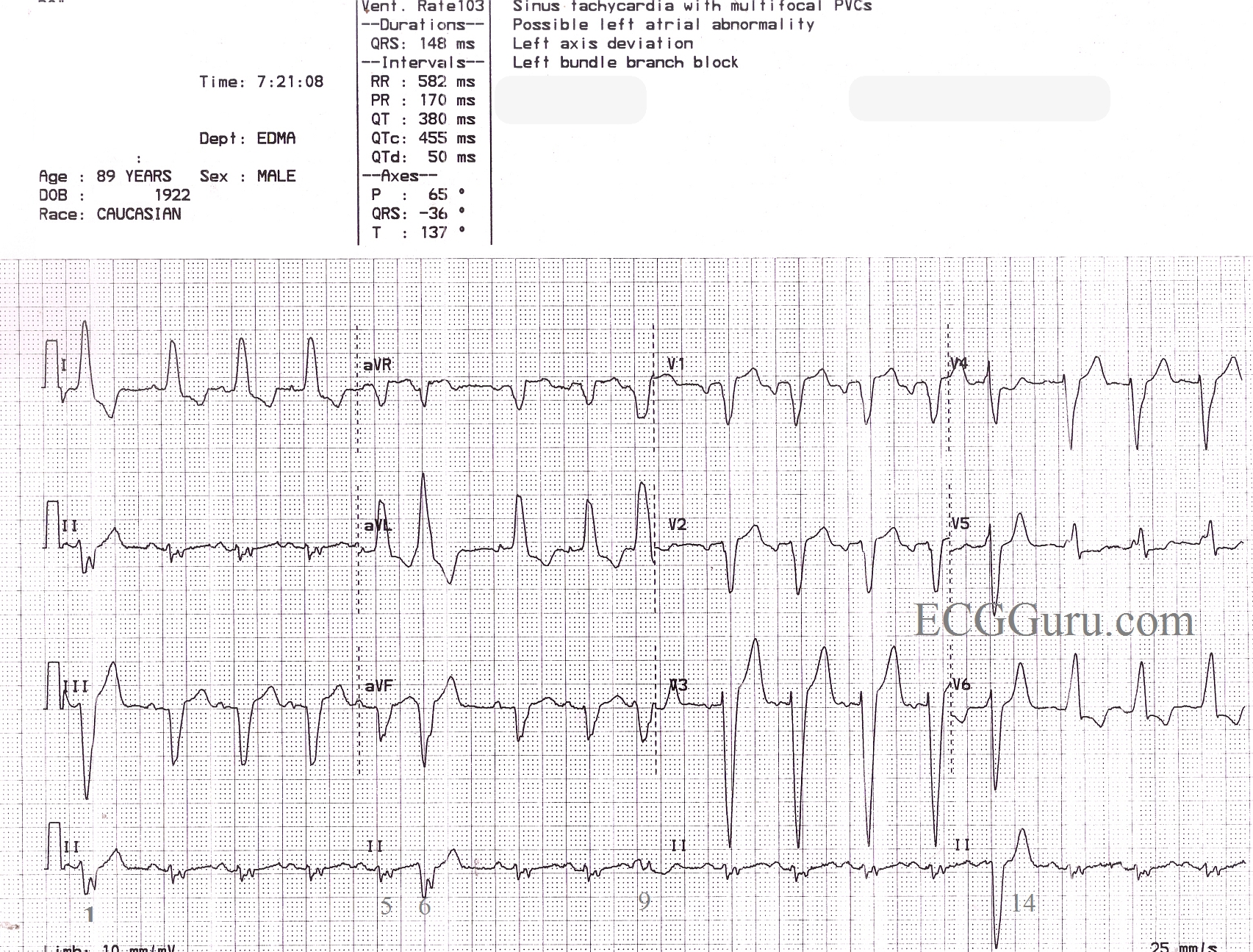
This is a great ECG for teaching your students about some of the different causes of wide QRS. This 89 year old man has a sinus rhythm that is around 100 bpm, and his QRS is widened at 148 ms (.148 sec). Leads I and V6 are positive, and Lead V1 is negative, meeting the criteria for left bundle branch block. There is a left axis deviation, which is common with LBBB, although it is not always this pronounced, indicating that there is possibly another cause for LAD. In this ECG, there are also PVCs and probable fusion beats. The 14th beat is a PVC. Complexes 1, 6, and 9 are possibly fusion beats. Fusion can be described as an almost simultaneous sinus beat and ventricular beat. The depolarization waves, one coming from the top of the heart and one coming from the bottom, meet and "fuse" on the ECG. Fusion beats will have some characteristics of the supraventricular beats and some of the ventricular beats. They are not significant except that fusion can be said to "prove" the existence of a ventricular pacemaker - either a natural pacemaker or an electronic one.
Do you see anything else interesting in this ECG? How would YOU describe this rhythm? Please do not hesitate to add your comments, or ask questions of the experts who contribute to this site. We will respond quickly to all questions.
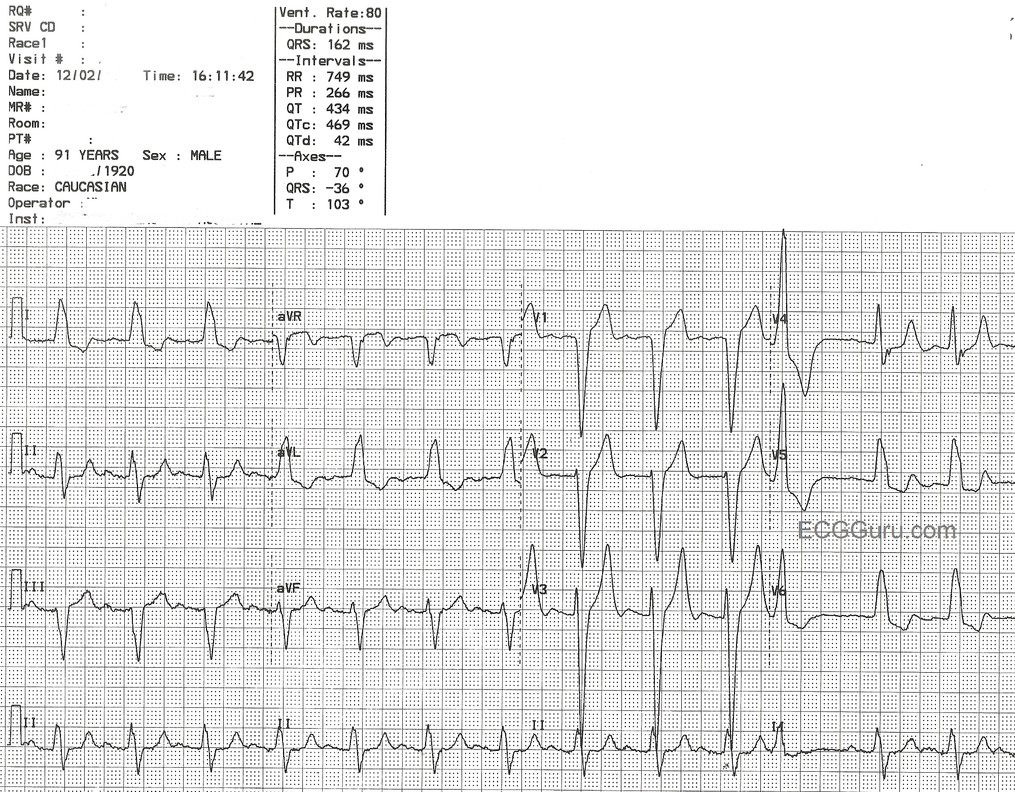
This ECG is from a 91-year-old man who was being evaluated for replacement of his aortic valve, which was severely calcified. It shows a classic LBBB pattern: wide QRS, supraventricular rhythm (normal sinus rhythm with first-degree AV block), a negative QRS in V1, and a positive QRS in Leads I and V6.