Dawn's picture

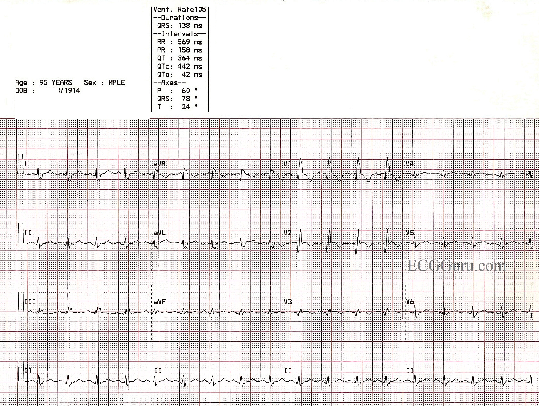
This is an ECG from a 95 year old man who was recovering from an anterior-septal wall M.I.  Other clinical data for this patient has been lost, except that he suffered a new right bundle branch block during this M.I.  The ECG shows pathological Q waves in V1, V2, and V3, consistent with permanent damage (necrosis) in the anterior septal wall.  The ST segments in those leads are coved upward.  Even though the J points are not elevated, this ST segment shape suggests recent injury.  The classic RBBB pattern is present:  wide QRS, rSR' pattern in V1, and wide little s waves in I and V6.  It is not known why the overall voltage is low in this patient.

Rate this content: 
Average: 4.3 (7 votes)

Comments

ekgpress@mac.com's picture

Great example by Dawn of RBBB with ECG signs of recent acute MI. I'll add several points to Dawn's description:

    Although it is more difficult to assess acute ischemia/infarction in the setting of QRS widening from BBB (Bundle Branch Block) - it will nevertheless be possible to do so in many cases. It is definitely easier to assess ischemia/infarction with RBBB than with LBBB. This has an anatomic & physiologic basis.

  • The initial part of the ventricles to be activation is the LEFT SIDE of the SEPTUM. Septal activation normally goes left-to-right. When there is LBBB - this direction of initial septal activation is altered (since the left bundle goes down the left side of the septum). Consequently - septal activation has to go from right-to-left when there is LBBB. You are therefore CHANGING the direction of the INITIAL ventricular activation with LBBB. Since Q waves are written during this initial ventricular activation period - detection of Q waves is far more difficult when there is LBBB ...
  • In contrast - the direction of initial ventricular activation is UNCHANGED when there is RBBB. This is because the right bundle branch goes down the right side of the septum. RBBB does not affect inital septal depolarization that still moves from left-to-right. As a result - you usually CAN see infarction Q waves with RBBB.
  • So it is with this tracing. We see large anterior Q waves (in V1,2,3) despite the RBBB. We have lost the rSR' in V1 (replaced with a QR) - but as per Dawn, we still recognize RBBB by QRS widening, the QR in V1 and the wide terminal S waves in lateral leads.
  • Had I not known the history in this patient - I would have interpreted this tracing as, "Sinus rhythm; RBBB; Anterior MI of uncertain age - possibly acute". The ST-T wave in lead V1 is consistent with RBBB and not particularly suspicious of acute infarction. But in addition to ST segment coving in leads V2,V3 - there IS suggestion of ST elevation in these leads (esp. in view of the fact that the ST segment should be BELOW the baseline slanting downwards if the only thing occurring was RBBB).
  • ST-T wave appearance is also distinctly abnormal in lead V4 - with coving and suggestion of recent injury (consistent with the ST-T waves in V2,V3).

Ken Grauer, MD  www.kg-ekgpress.com   [email protected] 

All our content is FREE & COPYRIGHT FREE for non-commercial use

Please be courteous and leave any watermark or author attribution on content you reproduce.