
Submitted by Dawn on Sat, 11/16/2013 - 21:26
We are continuing with the topic of teaching your students to use multiple leads to evaluate ECGs. Sometimes, beginners are given the impression that they need to learn 12-lead assessment only for the purpose of discovering ST elevation M.I. Of course, this is a very valuable use of 12-lead ECG, but when evaluating dysrhythmias and other cardiac issues, the more leads you have available to you, the more accurate you will be.
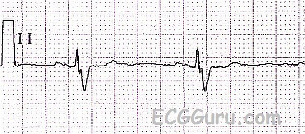
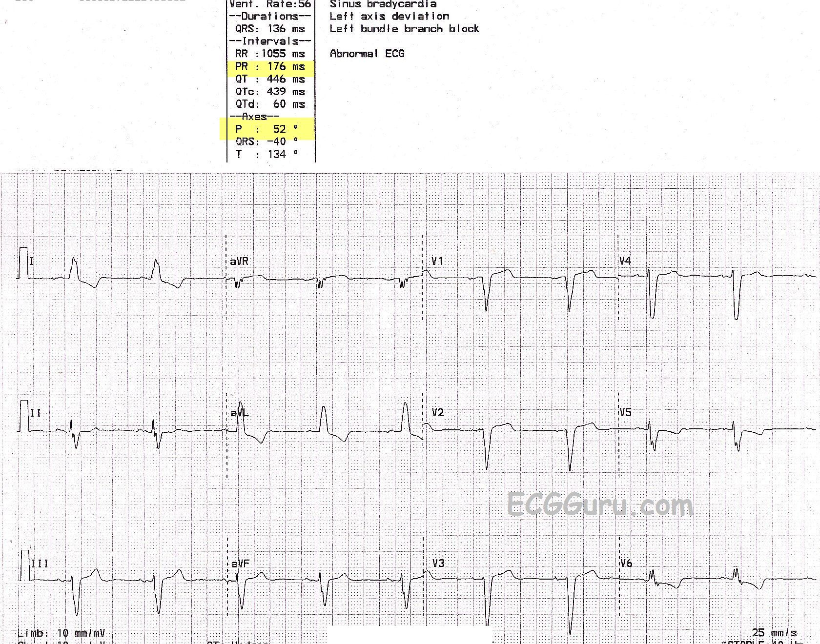
In today's example, the first short Lead II rhythm strip shows a sinus bradycardia. Your most experienced students may notice that the QRS complex is wide, at about .14 seconds. Most beginners, however, will fail to see this because the QRS is biphasic - partly positive and partly negative. This produces an illusion of a more narrow QRS. Show your students the same patient's 12-lead ECG, and they should readily see the wide QRS and the LEFT BUNDLE BRANCH BLOCK. It is difficult to demonstrate the criteria for LBBB without multiple leads.
To review, the ECG criteria for LBBB are: wide QRS, supraventricular rhythm, negative QRS in V1, positive QRS in Leads I and V6. In this example, the wide QRS is readily seen in monophasic leads such as Leads I and V1. The supraventricular rhythm (sinus brady) can be appreciated by seeing P waves in all limb leads except aVR, and the chest leads V3 through V6. If your students have difficulty determining the QRS width, have them look at the computer reading at the top (136 ms) and then check again with the QRS complexes. If P waves are difficult to see, look at the computer reading again. Did the computer measure a PR interval? In this case, yes. The PRI is 176 ms. The computer also provides a P wave axis. These are fairly reliable signs that P waves exist. Remember, in a 12-lead ECG, the multiple channels - in this case three - are set up to run simultaneously. Therefore, if a P wave is easy to see in one lead, but difficult to see in the same heartbeat above or below the easy one, we can assume the P wave exists in the hard-to-see lead. For example, in this ECG, we know V2 has a P wave because V3 does.
Try looking through some of the ECG archives here on the ECG Guru and see how many 12-lead ECGs you can find that would be misdiagnosed if only one lead was available.
Related Terms:
Rate this content:
All our content is FREE & COPYRIGHT FREE for non-commercial use
Please be courteous and leave any watermark or author attribution on content you reproduce.