Displaying 21 - 23 of 23
Dawn's picture

Inferior Wall M.I. With Third-degree AV Block

This ECG was obtained from an elderly woman who suffered a complete right coronary artery occlusion and inferior wall M.I.  In her case, the AV node was also affected, and she developed a third-degree AV block with a junctional escape rhythm.  A good ECG for ACLS classes as well as for ECG classes.  A lively discussion can be had regarding "types" of complete heart block and the nature of the escape rhythm - when to treat and when to leave the rhythm alone.  In this case the rate of the junctional escape rhythm was adequate for perfusion, and the patient's blood pressure was stable. Priority for treatment in this situation is restore blood flow through the coronary artery, if the patient is a candidate for PCI.  You might want to review Christopher Watford's contribution to the Ask the Expert page on AVB vs. AV Dissociation.

Dawn's picture

Third-degree AV Block (Complete Heart Block)

This 84-year-old man called 911 because he felt dizzy and fell.  He was not injured in the fall, but the paramedics noted a slow pulse. He denied significant medical history. The initial ECG showed sinus rhythm at about 80 bpm and AV dissociation with an apparent acellerated idioventricular rhythm at about 40 bpm.  Less than one minute later, he has developed a complete heart block with an idioventricular escape rhythm less than 30 bpm.  The escape rhythm speeds slightly toward the end of the strip.  He retained stable vital signs and adequate perfusion during transport.  It is presumed that he was scheduled for an implanted pacemaker.  It is interesting to note the machine's interpretation, and it reminds us to always interpret the ECG ourselves.   Thanks to ECG Guru member, Sebmedic, for his contribution of this ECG. 

Dawn's picture

Sinus Tachycardia With High-grade AV Block

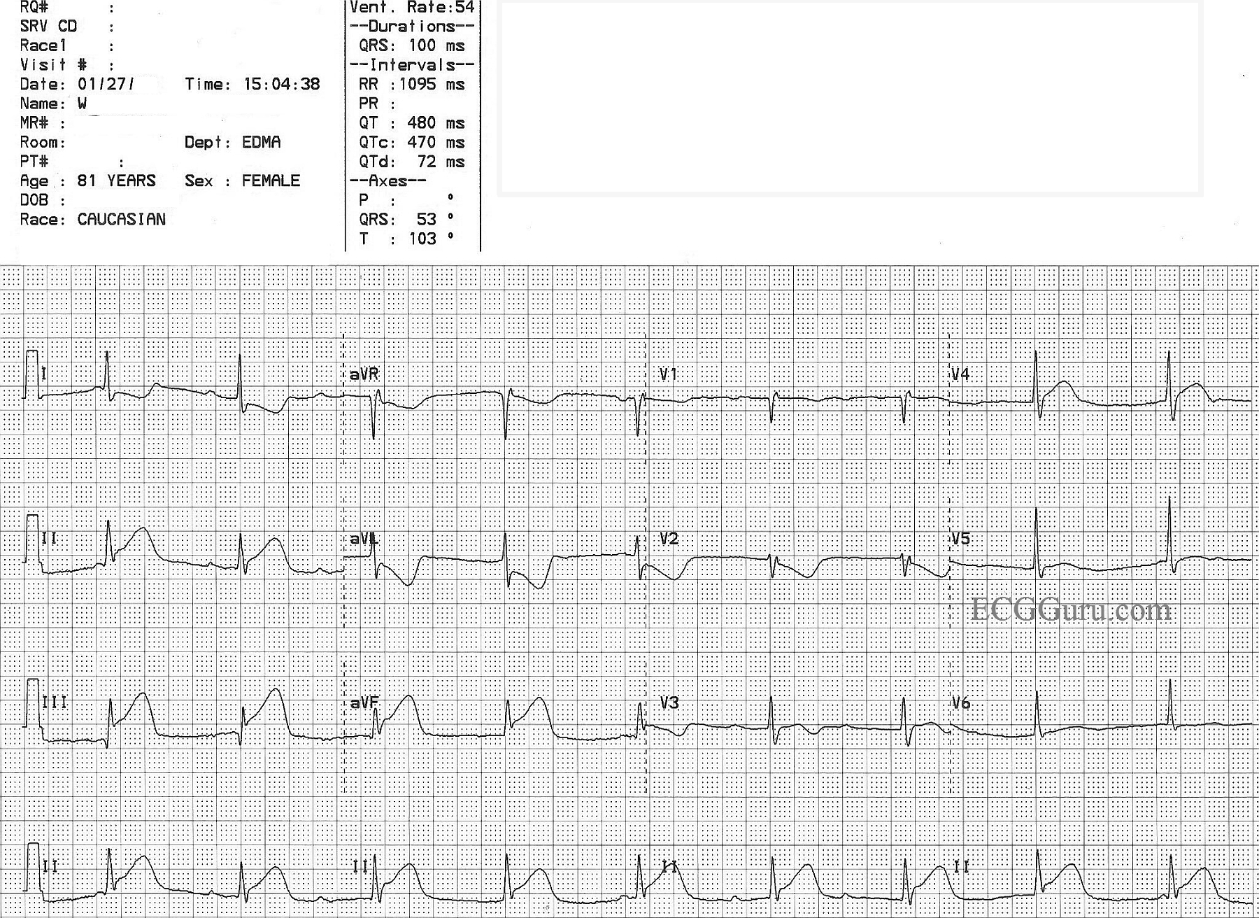
This ECG has always caused a lively conversation in ECG classes, both beginner classes and advanced. There is an obvious underlying sinus tachycardia, with clear P waves.

Some propose that the ECG shows a second-degree AVB, Type II, in that the PR intervals are constant, or nearly so. There is a slight discrepency if you compare the first PRI with the others. The QRS complexes, while very slow, have a pattern of right bundle branch block with left anterior fascicular block - not an unlikely finding in second-degree AV block, Type II, since that is a block in the fascicles of the interventricular conduction system. Type II blocks usually are accompanied by signs of bundle branch dysfunction.

Others strongly believe this is a third-degree, or complete, heart block. They argue that the PR intervals are not identical, and propose that a longer strip would uncover the discrepency. The wide QRS complexes have a strong left axis deviation, which could support the argument for idioventricular escape rhythm.

What do you think? Please comment below.

Pages

All our content is FREE & COPYRIGHT FREE for non-commercial use

Please be courteous and leave any watermark or author attribution on content you reproduce.