
Submitted by Dawn on Sun, 10/20/2013 - 22:29
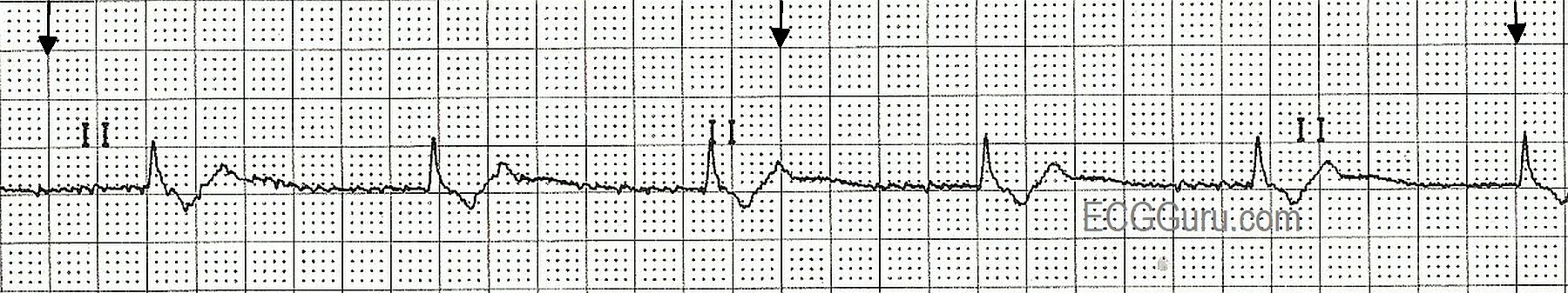
This rhythm strip illustrates a junctional escape rhythm. The sinus rhythm has slowed or stopped, and the junctional tissue has taken over as the pacemaker of the heart. The "junction" is loosely defined as the area between the AV node and the Bundle of His. The intrinsic rate of the pacemaking tissue in this area is 40 - 60 beats per minute. This slow rate is usually overridden by the sinus node, and the junction is not allowed to express itself as a pacemaker. Should the sinus node fail or fall below the junctional rate, the junction "escapes" and takes control of the heart. The QRS complex in junctional rhythm will normally be narrow, because the impulse follows the bundle branches down through the ventricles in a normal fashion, resulting in quick and normal ventricular depolarization. If the QRS complex is wide in a junctional rhythm, there is another, separate cause, such as bundle branch block.
If the junctional impulse is able to penetrate the AV node and depolarlize the atria, the P wave will be deflected downward in Leads II, III, and aVF, as the impulse is travelling in a retrograde direction (backward). The P wave could end up slightly before the QRS, during the QRS, or after the QRS. In this strip, the P waves are seen after the QRS complexes.
Rate this content:
All our content is FREE & COPYRIGHT FREE for non-commercial use
Please be courteous and leave any watermark or author attribution on content you reproduce.
Comments
Are You Certain this is a Junctional Rhythm?
I agree that the probable rhythm on this lead II rhythm strip is junctional at 55/minute. The reason I say "probable" - is that we ONLY see a single lead. There is a certain amount of baseline artifact on this tracing. Of relevance - is that the tail end portion of the QRS complex varies slightly from beat-to-beat and almost looks like its terminal portion is elevated above the baseline. Moreover - what is purported to be "retrograde P wave" almost blends in with this terminal part of the QRS. Bottom Line: I am not completely certain from this single lead tracing that the QRS complex is truly narrow - nor that the negative deflection that we see is truly a retrograde P wave vs the terminal portion of a wide QRS. To be CERTAIN - additional leads are needed. Part of the QRS may at times lie on or near the baseline in one or more leads - therefore simulating a narrow rhythm in some leads, when in effect the QRS is wide.
The SOLUTION is easy - Get more leads! Then you'll know for certain.
So - Once again touted "Basic ECG" - this is a tracing that could be educational for ANY level provider. IF I was teaching a basic provider - I would assure them that the QRS complex IS truly narrow in all 12 leads, and that we are only seeing one of those leads here. But if I was teaching a more advanced provider - I'd ask them the rhythm and follow up on whatever they said with the question, "Are you 100% certain?" If not - How could you become certain?
Final Point: As Dawn mentions - Junctional rhythms may show one of 3 types of atrial activity: i) negative P wave preceding the QRS in lead II; ii) no P wave at all in lead II; or iii) negative P wave after the QRS in lead II. Realize that the latter (ie, negative P wave after the QRS) is BY FAR the least common form of junctional rhythm. Most of the time - NO P wave at all will be seen. Sometimes you'll see a negative P preceding the QRS in lead II. It is rare that you'll see the situation suggested in this tracing.
Ken Grauer, MD www.kg-ekgpress.com [email protected]
Basic Level Example
This Lead II strip shows all the characteristics of junctional rhythm, as outlined in the original post. It is suitable for teaching the beginner student these criteria. Once your students learn multi-lead assessment and 12-lead interpretation, their horizons will broaden, and they will find that one lead is not enough. Without checking multiple leads, it is quite possible to mistake one rhythm for another, or to miss something very important. But, your students must walk before they fly. Learning the criteria of the basic dysrhythmias is a daunting task for the beginner, and having sample rhythm strips to illustrate these criteria is important. I personally feel that students should be taught from real ECGs from real people as much as possible. With that come some "grey areas" that must be handled sensitively by the instructor, taking into account the background and understanding of the student.
Dawn Altman, Admin