Dawn's picture

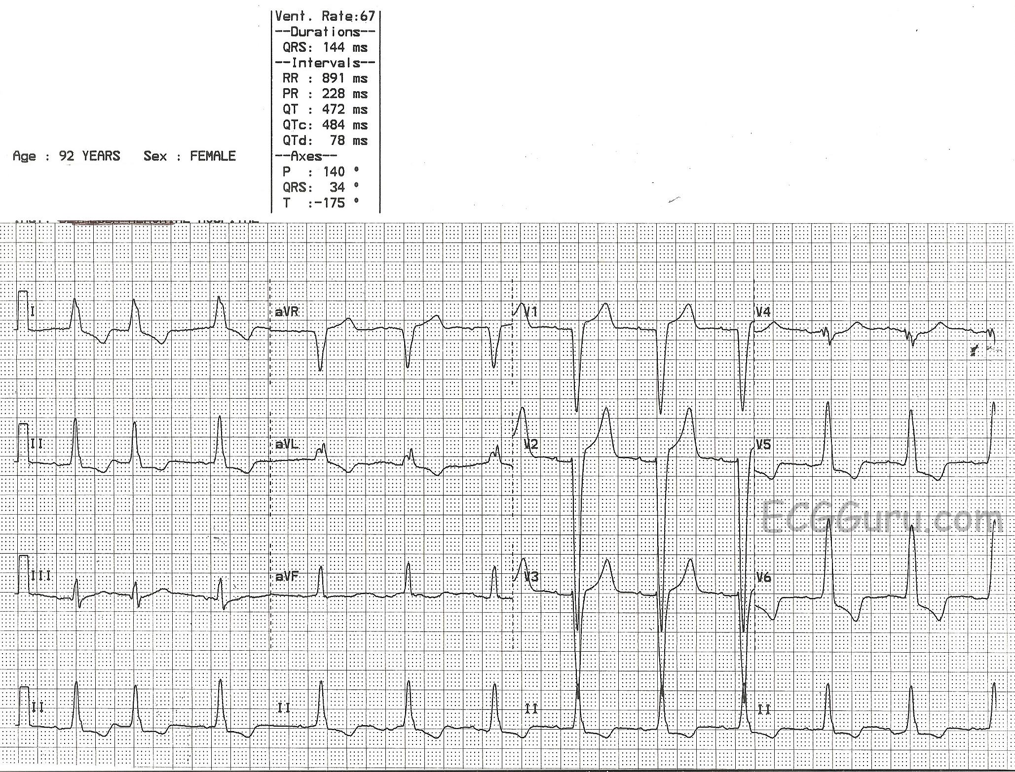
This is a good example of sinus rhythm with left bundle branch block.  There is some irregularity due to a PAC at the beginning.  The QRS is wide at 144 ms (.14 seconds).  There is also first-degree AV block, with a prolonged PR interval of 228 ms.  The criteria for diagnosis of left BBB are:  wide QRS, supraventricular rhythm, and a negatively-deflected QRS in V1 with a positive QRS in Leads I and V6.  

Left bundle branch block can be associated with many forms of heart disease, including CHF.  It can be permanent, transient, intermittent, or rate-related.  The wide QRS of LBBB significantly decreases cardiac output, causing poor perfusion symptoms in some people.

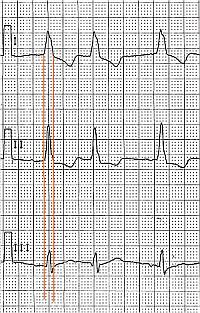
This ECG is a good one for your students who are just transitioning from reading rhythm strips to reading 12-lead ECGs.  It shows the value of multi-lead assessment of rhythms. You will notice that P waves are difficult to see in some leads.  Armed with the knowledge that the four channels on this ECG are run simultaneously, you can show the students how finding P waves in one lead will allow you to find them in the leads that are above and below that lead. 

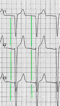
Similarly, it can be difficult to see the QRS width in some leads.  The leads in the same vertical column can help you see the QRS's true width, even if part of the QRS is "flat" in the isoelectric baseline.

Rate this content: 
Average: 5 (1 vote)

Comments

ekgpress@mac.com's picture

     As per Dawn - this ECG illustrates the appearance of complete LBBB (Left Bundle Branch Block). Review of all aspects of making the diagnosis of the bundle branch blocks (BBBs) extends beyond the scope of this short column - but I review this all in this BBB pdf excerpted from my ECG-2014-Pocket Brain. I'll limit my comments here to a few brief points:
  • The diagnosis of the BBBs can (and should) be made quickly. It generally takes us less than 5 seconds to confidently determine the type of BBB. The reasons this can be done so quicklly are: A) Because you only need to look at 3 leads ( = leads I,V1,V6) to diagnose BBB; and B) Because there are only 3 possible types we need to concern ourselves with = i) typical RBBB (Right Bundle Branch Block);  ii) typical LBBB; and iii) IVCD (IntraVentricular Conduciton Defect).
  • Typical RBBB is diagnosed by the presence of an rSR' complex in lead V1 with a taller right "rabbit ear" (ie, the R' is taller than the initial r wave). In addition - there are wide terminal S waves in lateral leads I and V6.
  • Typical LBBB is diagnosed by the presence of a predominantly negative complex in lead V1 (which may or may not have a small initial r wave) - and by a monophasic upright R wave in both leads I and V6. This is exactly what we see for the ECG presented in this case by Dawn.
  • IVCD is diagnosed when QRS morphology is not typical for either RBBB or LBBB in all 3 of the key leads (I,V1,V6). Again - this is all reviewed with pictures in the pdf I link to above.
  • ST-T waves present a predictable appearance when RBBB or LBBB is typical and there is no ischemia or infarction. Specifically - the ST segment and T wave are OPPOSITELY directed to the direction of the last QRS deflection. This is the case in this LBBB tracing - in which the ST-T wave is negative in leads I,V6 (ie, opposite the upright monophasic R wave in these leads) - and the ST-T wave is upright in lead V1 (ie, opposite the negative QS in lead V1).
  • QS complexes are common and expected with one or more of the anterior precordial leads with LBBB. We only see a QS in lead V1 here (a small r wave develops by lead V2) - but the point to make is that you cannot diagnose anterior infarction with LBBB even if QS complexes are seen in leads V1-thru-V4!
  • NOTE that there is at least 2mm of ST elevation in leads V1,V2,V3. This too is a normal finding with LBBB - especially given the upward concavity ("smiley"-shape) of the ST segment in these leads. This is NOT indicative of acute infarction. 
  • Finally - although LVH (Left Ventricular Hypertrophy) is difficult to diagnose in the setting of LBBB - it IS present in this case, because the anterior S waves are exceedingly deep (especially the S wave in lead V2, which is ~ 45mm deep). We presume LVH when there is LBBB if the S wave in V1 or V2 or V3 is 25-30mm deep - and it is much deeper here.

Ken Grauer, MD  www.kg-ekgpress.com   [email protected] 

All our content is FREE & COPYRIGHT FREE for non-commercial use

Please be courteous and leave any watermark or author attribution on content you reproduce.