Dawn's picture

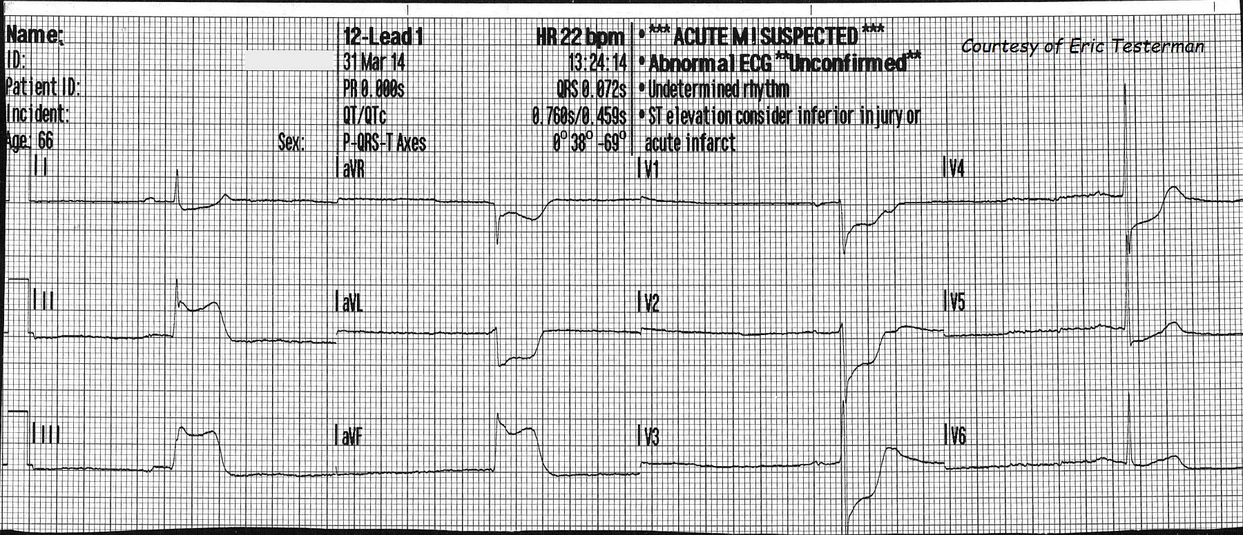
Another great ECG donated by Paramedic Eric Testerman.  This ECG is from a 66 year old man who was complaining of feeling dizzy, weak, and of having "minor" chest pain. He was extremely pale/ashen, had moderate cyanosis, and was very clammy and diaphoretic.  His initial heart rate was about 20 bpm.  His initial BP was 131/113 then, just before arrival at the hospital was 127/85. His HR increased to about 50 bpm (not shown). He was given 400 ml I.V. fluid, 324 gr of aspirin, and oxygen.  Transcutaneous defibrillator/pacemaker pads were applied. 

At the hospital, he was successfully treated with angioplasty for a 100% occlusion of the right coronary artery. The time from beginning of treatment to reperfusion of the artery was 47 minutes, which is very good! 

This is a "classic" inferior wall M.I., with ST elevation in leads II, III, and aVF. There are reciprocal ST depressions in I and aVL.  There are also ST depressions in V1 through V5.  This is generally considered to represent reciprocal ST changes in the posterior and lateral walls.There is a quite severe bradycardia, and the patient's skin showed signs of poor perfusion. Amazingly, the patient's BP stayed adequate during transport.  Bradycardia is common in inferior wall M.I. due to ischemic effects on the SA node and vagus nerve (sinus bradycardia) and the AV node (heart block).  In this case, the rhythm is sinus bradycardia.  The heart rate is in the 20's, and the PR interval is around .20 - .22 seconds. 

This patient's unstable condition and close proximity to the hospital prevented the EMS crew from getting a second ECG, but when there is time, a repeat ECG with a V4R lead can be helpful in deciding how much I.V. fluid is safe to give.  This patient's heart rate improved spontaneously during transport, but had it not, the transcutaneous pacemaker could have been used.  In some protocols, a trial of Atropine is advised.  In others, treatment goes straight to the pacemaker or to I.V. epinephrine.  During the angioplasty, a transvenous pacemaker is often inserted into the right ventricle to support the patient's rate if necessary.  

This patient was fortunate to be in an area with advanced paramedics and an interventional cath lab close by.  It is notable that he never developed pathological Q waves, and hopefully had an uneventful recovery.  


Rate this content: 
Average: 4.5 (2 votes)

Comments

ekgpress@mac.com's picture

     I support Dawn in appreciation for this ECG and case donated by Paramedic Eric Testerman - regarding this 66yo man with dizziness, chest pain, and a surprisingly reasonable BP ~120-130 mmHg systolic at the time of this and subsequent tracings. As per Dawn - there is severe bradycardia (rate in the 20's) and evidence of acute infero-postero MI. I'd add the following comments:
  • More than just "severe bradycardia" - I am not certain what the actual rhythm is. The R-R interval varies. P waves are of extremely small amplitude - and are NOT well seen in simultaneously-recorded leads aVR,aVL,aVF. I suspect there IS a P wave in these 3 leads - albeit the PR interval preceding the QRS in this lead appears much longer (~0.40 second) - so it seems that the PR interval is not constant throughout this tracing. I suspect the rhythm is actually either advanced 2nd degree or complete AV block - but a longer rhythm strip with P waves we can definitely see would be needed (in my opinion) in order to be certain of what the rhythm is.
  • Complicating our determination of what the rhythm might be - is ECG evidence of what I believe represents acute atrial infarction. This is best seen in lead III - where IF you look immediately after the P wave (and still just before the QRS in this lead) - you will see "TPa" elevation = elevation of the T component of the preceding P wave. This finding is also present in lead II, though somewhat less marked. I suspect that P wave with the long PR interval in lead aVR probably manifests reciprocal "TPa depression" - though clearly a better rhythm strip with more P waves of higher amplitude would be needed to be certain of this finding. However, atrial infarction DOES occur (it is usually overlooked ...) - it is most commonly seen in association with proximal RCA occlusion - and it may produce various subtle-but-real combinations of "TPa" elevation and depression in several leads. For more on Atrial Infarction - See Section 09.35 (beginning on Page 449) of this PDF excerpted from my ECG-2014-ePub on USING Lead aVR -
  • That there is almost certainly acute proximal RCA (Right Coronary Artery) occlusion can be gleamed from this ECG because of the following findings: i) Marked ST elevation in the inferior leads, with ST elevation in lead III > lead II; ii) Marked reciprocal ST depression, with ST depression more in lead aVL than lead I; iii) Suggestion of associated acute posterior MI with marked reciprocal ST depression in leads V1-thru-V4 and a slightly taller-than-expected R wave by lead V3 ("Positive Mirror Test" ); and iv) Lack of lateral precordial ST elevation, as might be expected if the patient had acute circumflex occlusion from a left-dominant circulation.
  • I wonder WHY Atropine was not tried? While use of this drug is not benign - this agent has by far BEST potential to be effective when used to treat Bradycardia due to an increase in vagal tone - something that is MOST likely to occur with acute RCA occlusion in the early hours of acute STEMI in association with narrow-QRS complex profound bradycardia and/or AV block. No more than 0.5 mg IV need be given initially, to see how/if the patient responds - but even though BP was 120-130 systolic, bradycardia (if not AV block) down to the 20's would seem indication for cautious trial of atropine (which might obviate need for pacing).
  • Finally - the questions arise as whether there is associated acute RV infarction? - and whether a lead V4R should be done to find out? Realizing there are several "correct answers" to this clinical question - I'd propose that a V4R lead should ONLY be done in the field IF it can be done in a matter of seconds (confirmed by the EMS team previously clocking with stopwatch the amount of time it takes them to obtain right-sided leads). Some set-ups allow immediate addition of right-sided leads to the ECGs obtained - others require extra time to do this. It is probably NOT worth doing ANYTHING in the field (like an extra set of ECG leads) if doing so takes delays transport by more than ~ 15-20 seconds because: i) We should presume RV involvement in cases of obvious proximal RCA occlusion such as this one - especially when there is significantly more ST depression in lead V2 compared to right-sided lead V1 (ST elevation from acute RV MI can be suspected when some of the expected ST depression in lead V1 is presumably cancelled out); and ii) Regardless of what a lead V4R might show - IV fluid should be cautiously given to a patient like this IF blood pressure drops (since relative hypovolemia is so commonly associated with these ECG findings).
--------------------------------
Additional LINKS of Interest:
  • For review of how to localize the site of Acute MI plus expected complications from the various infarct sites - Please check out my web page on this topic - CLICK HERE -
  • For Review on Use of Atropine (and other measures) in the Treatment of Bradycardia - CLICK HERE
 

Ken Grauer, MD  www.kg-ekgpress.com   [email protected] 

All our content is FREE & COPYRIGHT FREE for non-commercial use

Please be courteous and leave any watermark or author attribution on content you reproduce.