Displaying 21 - 30 of 31
Dawn's picture

Inferior Wall ST-elevation M.I.

This is an excellent example of acute inferior wall ST elevation M.I. (STEMI) in a 78 year old woman who had been experiencing intermittent chest pain for two days.  This 12-lead ECG has been formatted to simultaneously produce three rhythm strips below the 12-Lead.  The rhythm strips are run simultaneously with the 12-Lead, and show 3 different leads.  This feature can been invaluable when trying to decipher complex rhythms.

In this case, the patient is in normal sinus rhythm, and has classic ST segment elevation in the inferior wall leads:  II, III, and aVF.  There is some reciprocal ST depression in Leads I and aVL, and also in V1 and V2, suggesting injury extending up the posterior wall. ST depression is noticeable in V6 as well.  This patient's RCA lesion was opened and stented, and she did well immediately post cath.  We have no records past that.


Dawn's picture

Inferior Wall M.I. With Right Ventricular M.I.

This week's ECG of the Week is from an elderly woman who suffered an acute occlusion of the right coronary artery.  The ECG clearly shows ST elevation in leads II, III, and aVF, indicating inferior wall injury.  In this case, this ECG was obtained in the field by paramedics, and was the second ECG done on this patient. For this tracing, the paramedics obtained V3 and V4 on the right side to better view the right ventricle. V3 and V4 right clearly show ST elevation as well, indicating RVMI.  The slight coving and elevation observable in V1 is also an indication of RV involvement, and the ST depression in V2 indicates posterior wall injury.  All of this results from a proximal lesion of the RCA in this patient.  Such a lesion carries a high morbidity and mortality.

Taking the time to obtain a right ventricular lead is controversial in some settings.  Some believe the patient's hemodynamic condition should be treated, regardless of the presence or absence of ST elevation in right chest leads.  Others find it very helpful to know that the right ventricle is affected.  In this case, paramedics in this community have a protocol to avoid the use of nitroglycerine in RVMI patients, even when the BP is adequate.  So, for them, it is important to have the information gathered from V Right leads.

The rhythm here is interesting, as well, and not at all uncommon for IWMI patients.  The baseline artifact makes it a bit difficult to march out all the P waves, but it appears they are sinus P waves that are slightly irregular at a rate of 52 to 54.  The PR intervals appear to be progressively prolonging, but there is no "grouped beating" observable on this short strip.  A lack of a concurrent Lead II rhythm strip also makes it difficult to determine the rhythm, as Lead II does have very visible P waves, and would be helpful.  The regularity of the narrow complex bradycardia points to a junctional escape rhythm, which would make this a third-degree AVB at the AV node level, which is very common with IWMI.  What do you think?

This month's strip from Jason Roediger's ECG Challenge blogpost is a nice complement to the strip presented here.  His is much clearer, and has a Lead II rhythm strip.  Do you think these ECGs show the same rhythm, or two different rhythms?

For an excellent discussion of "AV Dissociation" vs. "AV Block", go to Christopher Watford's Ask the Expert post.  Thanks very much to ECG Guru Sebastian Garay for this interesting ECG.

Dawn's picture

Inferior Wall M.I. With Third-degree AV Block

This ECG was obtained from an elderly woman who suffered a complete right coronary artery occlusion and inferior wall M.I.  In her case, the AV node was also affected, and she developed a third-degree AV block with a junctional escape rhythm.  A good ECG for ACLS classes as well as for ECG classes.  A lively discussion can be had regarding "types" of complete heart block and the nature of the escape rhythm - when to treat and when to leave the rhythm alone.  In this case the rate of the junctional escape rhythm was adequate for perfusion, and the patient's blood pressure was stable. Priority for treatment in this situation is restore blood flow through the coronary artery, if the patient is a candidate for PCI.  You might want to review Christopher Watford's contribution to the Ask the Expert page on AVB vs. AV Dissociation.

Dawn's picture

Inferolateral M.I.

Unbelievably, this inferolateral ST elevation M.I. was missed by the treating paramedics in the field.  An elderly woman stepped off a curb and was hit by a very slow-moving car.  She fell and sustained a Colle's fracture of the right wrist. While the paramedics assessed her, she complained of chest pain, prompting them to perform a 12-Lead ECG.  The machine's interpretation called attention to the inferior and lateral walls' injury pattern, but the paramedics did not believe it, because "she was a trauma patient". They ran three ECGs, and still did not agree with the machine.

The patient was transported to a hospital without an interventional cath lab, and she was forced to endure a one-hour wait to be transferred to an appropriate hospital.

This is a great ECG for a discussion with your students about "distractors".  The call came in as a trauma, so that, in itself, was a distractor.  The rescuers saw what they expected to see. The angulated fracture distracted them - putting them into full trauma assessment mode.  Then, the frequent and coupled PVCs also distracted them, possibly making it more difficult for them to evaluate the ST segments in the normal beats.  Interestingly, the second and third ECGs did not have PVCs, and the ST elevation was even more clear.

PVCs which are repeating themselves in groups of two, three, or more are sinister in a chest pain patient, and may indicate LV dysfunction. They could possibly result in ventricular tachycardia, which would be disasterous for this patient.

Dawn's picture

Inferior Wall M.I.

In this ECG, there is ST elevation in II, III, and aVF and reciprocal ST depression in I and aVL, indicating acute inferior wall M.I. Also, this patient has developed pathological Q waves in III and aVF, and probably II as well, indicating permanent damage to the myocardium.  A ventriculogram or echocardiogram will confirm akinesis of the inferior wall in most cases.  In this ECG, there is no ST depression in V1 through V3, so we can hope the posterior wall has been spared.  Notice the flattening of the ST segments in the elevated leads.  This is a sign of CAD.  Last week's ECG had coved upward (frowning) ST segments, which are even more sinister looking.  Lead V1 has the flattening, and a pathological Q wave.  When V1 looks "sick" and V2 looks "well", there is a good probability of right ventricular injury as well.  Lead III has a taller ST segment than Lead II.  This has also been shown to be a marker of RVMI.  Regardless, a right-sided ECG, or at least a V4 right, should be obtained in any IWMI, since the RCA often supplies both the right ventricle and the inferior wall of the left ventricle.

The rhythm in this ECG is interesting, as well.  It appears to be sinus, but it is difficult to evaluate P waves.  In the Lead II rhythm strip, they appear to change in morphology. Because the R to R interval remains constant, we feel this change in appearance is due to baseline artifact caused most likely by patient movement such as breathing.  What do you think?  

Our thanks to Andrew Porter for contributing this ECG.

Dawn's picture

Inferior Wall M.I. With Sinus Bradycardia and First-degree AV Block

Inferior wall MI: ST elevation in II, III, and aVF. Reciprocal ST depressions. Sinus bradycardia and first-degree AV block suggests sinus node and AV node ischemia. This is a good "classic" inferior wall M.I. It is good for teaching inferior-posterior injury, and the effects of RCA occlusion on the sinus and AV nodes. The low voltage in the limb leads may also be due to acute M.I., but in this case, we do not know the patient's body size.


Dawn's picture

Atrial Pacing in a Patient With Acute Inferior Wall M.I.

Some people have been taught (incorrectly) that an electronic pacemaker prevents us from seeing an acute ST elevation M.I.  Not true.  It can be difficult to interpret ST elevation M.I. in the setting of WIDE QRS complexes.  When this situation exists, it is best left to the experienced ECG interpreter to determine whether there is STEMI.  In this ECG, we see ATRIAL pacing.  The patient has an intact AV conduction system.  The pacemaker paces the atria, and the impulse continues normally through the AV node and the ventricles.  The QRS that results is normal (narrow).  In this situation, the ST segments are accurate for determining ST elevation and depression.

This patient is a 74-year-old man who complained of chest pain for five days before presenting to his primary physician at the outpatient clinic.  After obtaining this ECG, the PCP transferred his patient to the Emergency Department.  He was admitted to the CCU with troponin level of 2.13 ng/ML. (Normal < 1.5 ML)


Our thanks to Jason Roediger, ECG GURU, for contributing this ECG.


Dawn's picture

AWMI With Recent IWMI

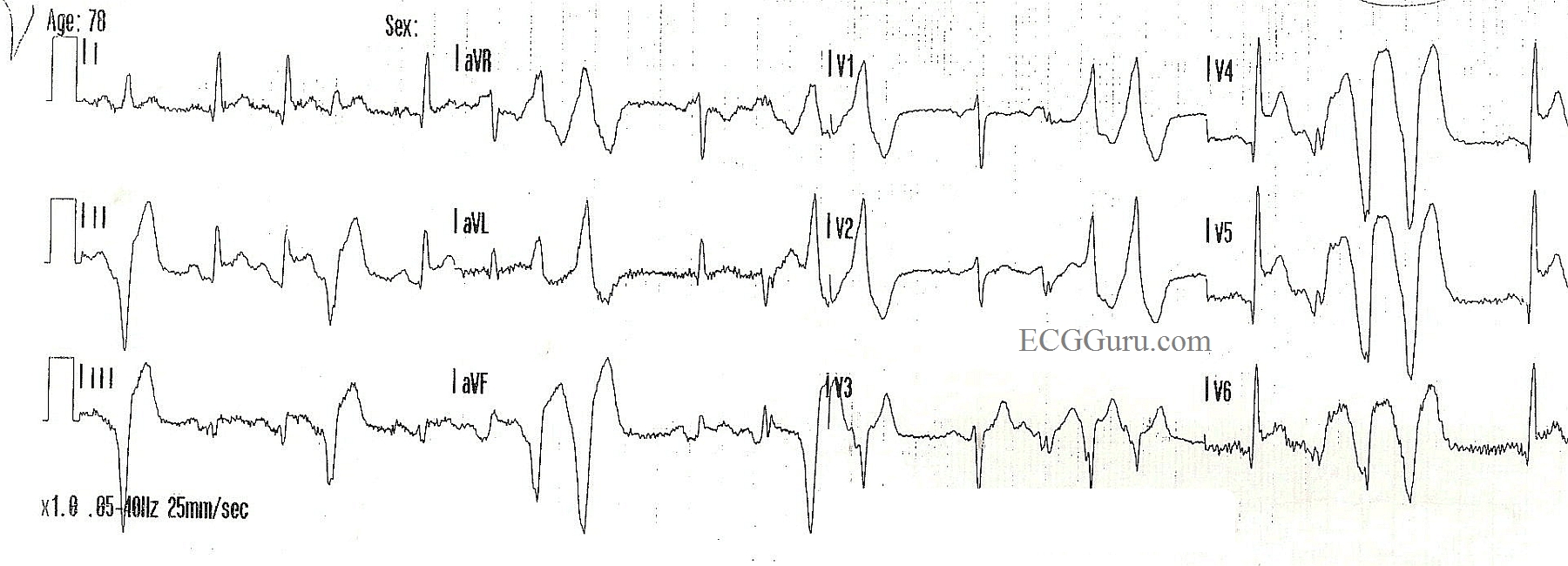
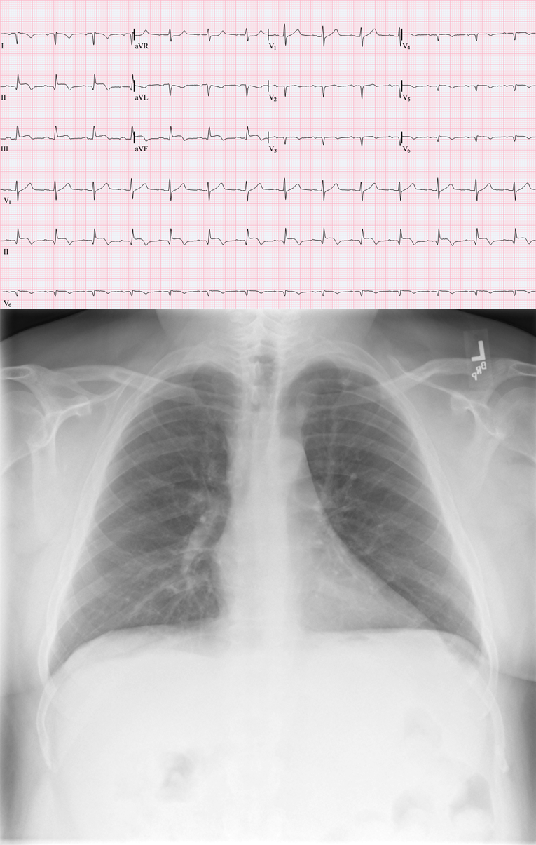
Patient AW103: This 88 year old woman had been sick for several days, but had not sought treatment. Her family found her nearly unresponsive and called 911. She presented to the Emergency Department as a STEMI Alert, and was in cardiogenic shock, with very poor perfusion. The ECG from the ED shows a large antero-lateral M.I., with ST elevation in V2 through V6, and also I and aVL. In addition, there are pathological Q waves, indicating necrosis, in the precordial leads, V2 through V6. The inferior wall leads, II, III, and aVF, also have pathological Q waves and abnormally shaped ST segments - no longer distinctly elevated, but coved upward.

This is a good tracing to teach students about Q waves and "old", "new", and "recent" M.I., and also about the clinical effects of hypokinesis or akinesis of the ventricles.

Unfortunately, this patient suffered a cardiac arrest in the cath lab while having her LCA reperfused with balloon angioplasty. She was resuscitated, on a ventilator and intraaortic balloon pump, and admitted to the CVICU, where she passed away within a few hours.

You will find photos from her cardiac cath in the Other Instructor Resources section, labelled as patient AW103. Click here for RCA Image, LCA Occluded Image, LCA Angioplasty Image.  Videos of her ventriculogram and left coronary artery angiogram can be found in the Resources section of this website.

Dawn's picture

Inferior - Posterior M.I.

This ECG shows a classic inferior - posterior STEMI.  This M.I. was due to complete occlusion of the right coronary artery.  ST elevation apparent in Leads II, III, and aVF show the acute injury in the inferior wall, while ST depressions in V1 and V2 are reciprocal of the ST elevations in the posterior wall.  The tall R waves in Leads V1 - V3 most likely are reciprocal to pathological Q waves in the posterior wall.  Tall R waves in the right precordial leads can be caused by other cardiac conditions, such as right ventricular enlargement.  RV hypertrophy can probably be ruled out in this case because there is no right axis deviation or P pulmonale.  Because inferior wall M.I.s often extend into the posterior wall, it is the most likely cause of the tall R waves.

Pages

All our content is FREE & COPYRIGHT FREE for non-commercial use

Please be courteous and leave any watermark or author attribution on content you reproduce.