
Submitted by Dawn on Tue, 12/16/2014 - 00:00
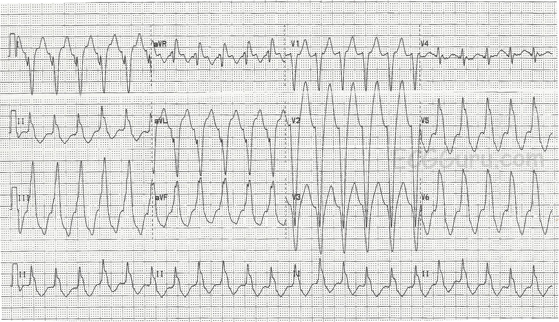
This ECG was taken from a patient who was complaining of palpitations and tachycardia, but who was hemodynamically stable, with no history of heart disease. It is an example of RIGHT VENTRICULAR OUTFLOW TRACT TACHYCARDIA, a type of idiopathic ventricular tachycardia. The ECG signs of RVOT are: wide QRS complex, left bundle branch block pattern (QRS negative in V1 and positive in Leads I and V6), heart rate over 100 bpm, rightward or inferior axis (LBBB usually has a normal to leftward axis), AV dissociation.
RVOT accounts for about 10% of all ventricular tachycardias, and 70% of idiopathic VT. It is most often found in structurally normal hearts, but it may occur in patients with arrhythmogenic right ventricular dysplasia. For more on RVOT, read Life In the Fast Lane.
RVOT tachycardia sometimes converts with adenosine. The patient in this example converted after being administered amiodarone.
Related Terms:
Rate this content:
All our content is FREE & COPYRIGHT FREE for non-commercial use
Please be courteous and leave any watermark or author attribution on content you reproduce.