
Submitted by Dawn on Sat, 06/02/2012 - 14:02
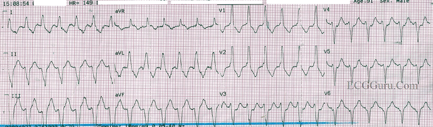
This wide complex tachycardia occurred in a 91 year old man with a history of atrial fibrillation. He complained of "fluttering" in his chest, and denied chest pain or other problems. While the paramedic attempted to start an I.V., he spontaneously converted to atrial fibrillation with left BBB, and PVCs. Once he converted, his symptoms abated. Remember, all wide complex tachycardias (WCT) should be treated as V Tach in the field, as this is by far the most common WTC and the most dangerous.
Some of the ECG clues that this WTC is ventricular tachycardia are:
* Monophasic upright QRS in V1 (does not have RBBB pattern of rsR')
* Extreme left axis deviation (II, III, and aVF are negative, I, aVL, and aVR are poisitive)
* V6 is negative
For a more thorough discussion of the ECG signs of V Tach, go to Jason Roediger's Ask the Expert page discussion on the topic:
Rate this content:
All our content is FREE & COPYRIGHT FREE for non-commercial use
Please be courteous and leave any watermark or author attribution on content you reproduce.
Comments
Certainly VT - Perhaps Belhassen Tachycardia
A positive aVR certainly seals this as VT. However, interestingly enough the QRS duration isn't quite as wide as we normally would believe to be the case in VT. Given this is V1-positive with an extreme Left Axis Deviation, this could be a case of Belhassen Tachycardia or an Idiopathic Left Posterior Fasicular Tachycardia. Cardiologists may choose to treat this with verapamil, however, an incorrect diagnosis on our part could cause VF.
In the field treating this as VT is the only safe bet!
Christopher
sixlettervariable.blogspot.com
ems12lead.com
re: agree with VT
Regular monomorphic WRST wih cycle length 400 ms (150/min), RBBB (predominantly monophasic R), left superior axis, positve aVR, transition at V3, R/S < 1 in V6, RS (V4 ) about 100 ms and AVD. Findings suggestive of VT. I thought that aVR is narrow but looking at synchronous leads aVF, the terminal component of aVR is part of the QRS. I also event thought that was an F wave. RBBB, Left superior axis, S in V6 and pronounced R in aaVR suggest what was mentioned by Christopher as L post fascicular VT.