This image is taken from the patient featured in our Teaching Series, AWMI. This shot shows the left anterior descending artery just after a stent has been placed and blood flow returned. There had been a 100% occlusion prior to the angioplasty. The circumflex and obtuse marginal arteries show evidence of severe coronary artery disease and multiple partial obstructions, and were also repaired.
This is an original illustration by Dawn Altman. It is free for your use in an educational setting. For other uses, please contact Dawn at [email protected].
Looking for an illustration for your presentation or packets?
Photograph by Alyssa Thompson. May be used free of charge and free of copyright in instructional setting. Please contact the artist at [email protected] for any commercial use.
An angiogram of the left coronary artery of a patient with multi-vessle coronary artery disease. Pacemaker electrodes are seen in the right atrium and right ventricle. A Swan Ganz catheter is also visible, with balloon inflated.
This image is courtesy of Dr. Stasinos Theodorou of the Limassol Cardiology practice. It may be used free of charge and free of copyright for educational purposes. For any commercial use, please contact Dr. Theodorou at the Limassol Cardiology practice.
This is a right coronary artery that was 100% occluded in the distal portion, and has now been successfully repaired. This photo is courtesy of Dr. Stasinos Theodorou of the Limassol Cardiology practice, and may be used free of charge and free of copyright for educational purposes. For any commercial use, please contact Dr. Theodorou at http://www.cardiolimassol.com
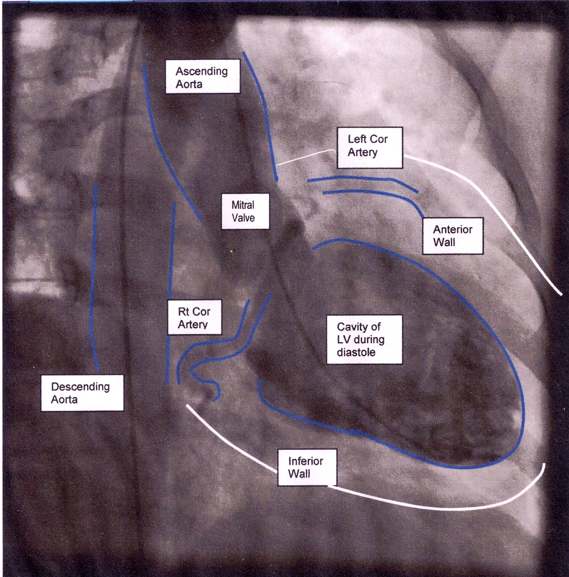
This image is courtesy of Dr. Stasinos Theodorou of the Limassol Cardiology practice. It may be used free of charge and free of copyright for educational purposes. Any commercial use would require permission from Dr. Theodorou: http://www.cardiolimassol.com.
The ECG Guru now has a You Tube site where you can find videos to enhance your classes. As with all ECG Guru content, there is no charge and no copyright. Ventriculograms are often obtained during a cardiac cath procedure. A curved, or pigtail, catheter is inserted through the arterial access line into the aorta and then the left ventricle. Contrast is introduced into the left ventricle and the pumping function of the ventricle can be observed. The structure and function of the aorta and mitral valve may also be observed during this procedure.