The Patient:An elderly man presents with chest pain, pallor, diaphoresis and weakness.
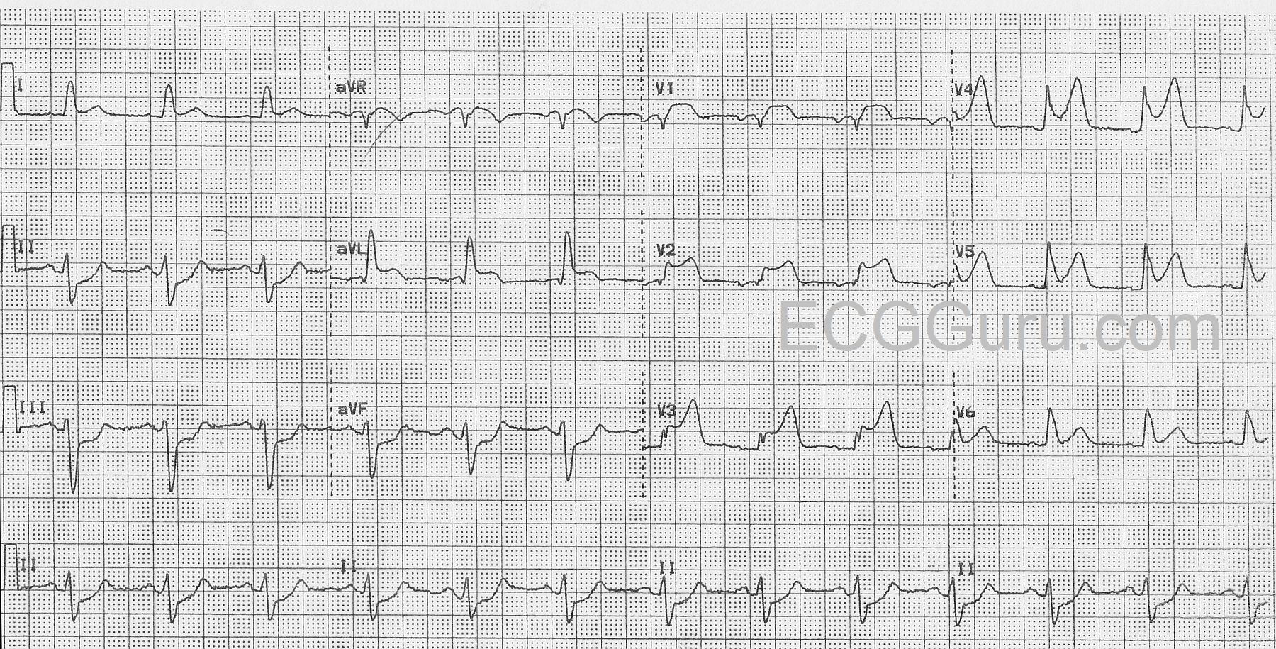
The ECG:The rhythm is normal sinus at a rate of about 76 bpm with normal intervals. The QRS complexes are wide at about .14 seconds (140 ms).There is ST segment elevation in all precordial leads, except for possibly V6.The shape of the ST segments in the anterior wall range from coved upward in a “frowning” shape (V1) to very straight (V5 and V6).There is also ST elevation in aVL with ST straightening in Lead I.There is ST depression in the inferior leads, II, III, and aVF.Lead II is equally biphasic while I and aVL are positive, indicating an axis that is shifted slightly to the left.With his symptoms and this alarming ECG, he was sent promptly to the cath lab.
Interpretation:The rather obvious ST-elevation M.I. is extensive, covering the entire anterior wall, and extending into the high and low lateral walls . This was confirmed in the cath lab, as the patient had an occlusion of the left anterior descending artery near the bifurcation of the circumflex.The wide QRS meets the criteria for left bundle branch block (wide QRS, negative QRS in V1 and positive QRS in V6 and Lead I).However, it doesn’t have the “look” of LBBB with the low-voltage seen in the anterior wall. After the offending artery was opened and stented, the wide complex became narrow and was considered to be an interventricular conduction delay that was due to the ischemia.The ST depression in the inferior wall is most likely reciprocal.