
Submitted by jer5150 on Sun, 08/19/2012 - 20:19
Patient clinical data: 68-year-old black man.
Question:
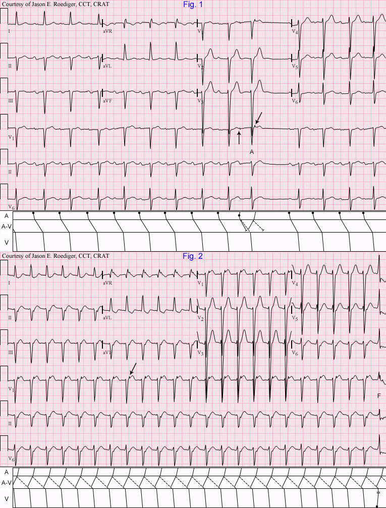
(1.) What "pseudo" clue in Fig. 1 clinches the source of the mechanism seen in Fig. 2?
Rate this content:
-

- jer5150's blog
- Log in or register to post comments
All our content is FREE & COPYRIGHT FREE for non-commercial use
Please be courteous and leave any watermark or author attribution on content you reproduce.
Comments
A Pseudo-R' in V1 and Pseudo
A Pseudo-R' in V1 and Pseudo-S in II (although II already has a true S from LAFB) are suggestive of AVNRT, coupled with the PAC in first strip make that mechanism for the tachycardia seem more likely.
Christopher
sixlettervariable.blogspot.com
ems12lead.com
I echo what Christopher said,
I echo what Christopher said, but also have to point out the pseudo-R' in V1 of the QRS following the PAC, clinching the diagnosis of a reentry circuit.
Vince D
http://www.medialapproach.com
I Re-Echo VinceD ( "pun" fully intended )
Christopher & VinceD are right on the mark. Jason soon coming with a gorgeous laddergram. I'd just add its always nice when you can see evidence of reentry (retrograde conduction back over the fast pathway producing notch in terminal part of the QRS) in many leads. In the 2nd tracing - in addition to leads V1 & lead II - we also see that retrograde P in leads III,aVR,aVF,V5 and V6 - which as per Vince "clinches" the diagnosis of reentrant SVT.
Ken Grauer, MD www.kg-ekgpress.com [email protected]
INTERPRETATION
(1.) Sinus rhythm (rate about 82/min) with . . .
(2.) . . . so-called “first-degree” A-V block (P-R = 0.26s).
(3.) Left axis deviation (LAD; about -59 degrees) probably due to . . .
(4.) . . . left anterior hemiblock (LAHB).
(5.) One atrial premature beat (APB; 10th beat) with prolonged P’-R interval .
(6.) One atrial “echo” (i.e., reversed reciprocal beat) producing “pseudo r’-wave” (oblique arrow).
Interpretation: (Fig. 2)
(1.) Atrioventricular nodal reentrant tachycardia (AVNRT) at a rate of about 143/min.
(2.) One probable ventricular fusion beat (F; 24th beat)
(3.) Left axis deviation (LAD; about -66 degrees) probably due to . . .
(4.) . . . left anterior hemiblock (LAHB).
Comments:
Many thanks to Christopher, Vince, and Ken for participating in this week’s case study. There is very little left for me to contribute to their respective answers. Both of these serial ECGs were recorded in separate, consecutive years. In the laddergram's A-V tiers, the solid lines (i.e., ––––––––– ) represent conduction via the fast pathway (FP) and the broken / dashed lines (i.e., - - - - - - - - ) represent conduction via the slow pathway (SP).
Jason E. Roediger - Certified Cardiographic Technician (CCT)
[email protected]
AVNRT
The pseudo R prime in V-1 during the tachycardia that is not present before the tachycardia is not only suggestive
of slow-fast AVNRT, it is diagnostic of it. In a large EP study, this was 100% diagnositic of slow-fast AVNRT.