
Submitted by jer5150 on Sun, 05/18/2014 - 10:12
Patient data: 54-year old man who recently underwent a major cardiac procedure. At first glance, this ECG may not appear to be particularly unique but a closer inspection reveals something unusual going on here.
HINT: The telling clue can be found in lead II but you'll have to use calipers and think "outside-the-box". 3 days after this ECG, the patient reverted back to sinus rhythm but continued to display this one finding. Good luck!
Spoiler alert!!!
Only click on these 2 links if you want the answer given to you.
Link 1
Link 2
Rate this content:
-

- jer5150's blog
- Log in or register to post comments
All our content is FREE & COPYRIGHT FREE for non-commercial use
Please be courteous and leave any watermark or author attribution on content you reproduce.
Comments
A-flutter with a 4:1
A-flutter with a 4:1 conduction rate.
Awesome!
I'm not going to give away the answer just yet since no one else has identified it, but this is an absolutely awesome tracing Jason! Good eye for picking this up!
Vince D
http://www.medialapproach.com
Thanks!
Thank you for the compliment Vince. This is one of my favorite ECGs in my collection!
Jason E. Roediger - Certified Cardiographic Technician (CCT)
[email protected]
Curious EKG
Atrial Fib with a narrow QRS complex with constant R-R and frecuency of around 75bpm. I will consider hisian pacing, left pacing, or even stranger one atrium with a fib and the other (right) in sinus rhythm.
intermittent pre-excitation?
Could there be a delta wave in beats 2 and 8 or is it just my imagination?
Dave R
Too difficult for me!
I've just checked out the 'spoilers'. I would never have got this one!
Dave R
In all honesty . . . .
In all honesty, I may not have recognized this either had I not reverse-engineered my interpretation. It was only after I looked at the patient's ECG showing sinus rhythm that I thought to go back and look at some of their other recent ECGs and that's when I saw what was happening here.
Jason E. Roediger - Certified Cardiographic Technician (CCT)
[email protected]
INTERPRETATION
INTERPRETATION:
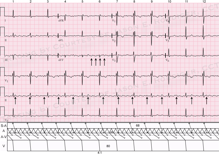
(1.) Atrial flutter (rate about 300/min) with 4:1 A-V conduction reducing the ventricular rate to about 75/min (see laddergram).
(2.) Incomplete right bundle-branch block.
(3.) Sinus rhythm at a rate of about 87/min (best seen in lead II).
COMMENTS:
Status-post orthotopic heart transplant. The donor heart is in atrial flutter (arrows in lead aVF) and the atrial cap of the recipient heart is in sinus rhythm. Note that the sinus P-waves can be seen marching through and deforming the sawtooth pattern of the flutter waves (arrows in lead II). In the laddergram, the sinoatrial (S-A) teir is meant to represent the S-A node of the recipient heart's atrial cap. As I previously mentioned, the patient's donor heart reverted back into sinus rhythm 3 days later. At that time the donor heart was in sinus rhythm at a rate of 83/min and the atrial cap of the recipient heart was in sinus rhythm at a comparable rate of 79/min.
This same scenerio could be seen in a heart with dissociated atrial rhythms. The right atria in sinus rhythm and the left atria in atrial flutter.
Jason E. Roediger - Certified Cardiographic Technician (CCT)
[email protected]