Displaying 11 - 19 of 19
Dawn's picture

ECG Basics: Atrial Pacing

This strip for your basic students is a nice example of atrial pacing in a patient with an intact interventricular conduction system.  Generally, the pacemaker will behave this way when the sinus node is not functioning well enough to provide adequate rate for the patient, and the conduction system from the AV node down is functioning properly.

Pacemakers in the modern age are very complicated to understand for the beginner, and pacemaker programming and malfunctions often cannot be determined from a simple rhythm strip.  It can be a challenge to teach beginning students about the programming options available today.  This strip is nice because it is clear, and the pacer spikes are readily seen.  The patient is being paced 100% of the time in this strip.


jer5150's picture

Jason's Blog: ECG Challenge of the Week for Sept. 23-30, 2012.


Patient's clinical data:
75-year-old white man who presented to the emergency department.  The patient was ventricularly paced on an emergent basis and the indication was probable complete AV block with an ineffective junctional escape rhythm.  I don't believe this patient survived the admission of this hospital visit.    

Dawn's picture

AV Sequential Pacing to Ventricular Tachycardia

This is an interesting ECG for showing students AV sequential pacing and also ventricular tachycardia.  The unusual thing about this ECG is that the V Tach starts at the time the machine begins recording the precordial leads.  This particular ECG machine shows a slight "gap" at the lead change, so we don't see the actual start of the V Tach.  Both rhythms have wide QRS complexes. The pacemaker is pacing the right ventricle, so you will see a wide QRS with a leftward axis, as the impulse spreads up and leftward toward the left ventricle.   The V Tach portion is, of course, limited to the precordial leads, so we cannot plot the frontal plane axis.  But, it meets many of the accepted criteria for ventricular tachycardia, including:  very wide QRS, negative QRS in Lead V6, absence of RBBB or LBBB pattern.   For more on recognizing V Tach in a WCT, go to Ask the Expert at this LINK.    

This is also a very good example of how the interpretation by the machine can be wrong.  Always read the ECG yourself!

Dawn's picture

Atrial Pacing in a Patient With Acute Inferior Wall M.I.

Some people have been taught (incorrectly) that an electronic pacemaker prevents us from seeing an acute ST elevation M.I.  Not true.  It can be difficult to interpret ST elevation M.I. in the setting of WIDE QRS complexes.  When this situation exists, it is best left to the experienced ECG interpreter to determine whether there is STEMI.  In this ECG, we see ATRIAL pacing.  The patient has an intact AV conduction system.  The pacemaker paces the atria, and the impulse continues normally through the AV node and the ventricles.  The QRS that results is normal (narrow).  In this situation, the ST segments are accurate for determining ST elevation and depression.

This patient is a 74-year-old man who complained of chest pain for five days before presenting to his primary physician at the outpatient clinic.  After obtaining this ECG, the PCP transferred his patient to the Emergency Department.  He was admitted to the CCU with troponin level of 2.13 ng/ML. (Normal < 1.5 ML)


Our thanks to Jason Roediger, ECG GURU, for contributing this ECG.


Dawn's picture

Pacemaker Rhythm, Right Ventricular Pacing Triggered by Native P Waves

This patient's electronic pacemaker has pacing electrodes in the right atrium and the right ventricle, which is typical.  In this ECG, the patient produces P waves, but fails to transmit them to the ventricles due to AV block.  The electronic pacemaker SENSES the native P waves, and TRIGGERS the ventricular wire to pace the ventricle in response.  Using this pacemaker function, the heart rate can be controlled by the patient's own nervous system, as P waves are produced naturally.  Should the patient fail to produce a P wave within rate guidelines set by the implanting physician, the pacer will pace the atrial as well.

The QRS complex is wide when only the right ventricle is paced, as the impulse has to travel from the RV to the LV cell-by-cell.  This slows conduction in the same way a ventricular rhythm like VTach is slowed, producing a wide QRS.  Remember, a wide QRS causes lower cardiac output, as much as 15%.

For your students who are learning axis, show them an illustration of the heart with the pacing wire in the right ventricle.  It is easy to see why the axis is leftward when the left ventricle is being paced from the right ventricle.

Dawn's picture

Pacemaker Mediated Tachycardia

PACEMAKER MEDIATED TACHYCARDIA is a term for several different mechanisms that cause inappropriately fast rates in a paced patient. It is a very interesting topic, which I will not attempt to discuss here, because it has been done to perfection by DR. KEN GRAUER, MD, with assistance from JASON ROEDIGER, CCT, CRAT in Dr. Grauer's ECG Consult #13 Please follow this link to a concise, complete, and illustrated discussion of PMT.

Dawn's picture

Pacemaker Mediated Tachycardia

    Do you ever feel confused by pacemaker rhythms?  You are not alone!   Pacemakers are electronic devices, and have rapidly evolved in their capabilities.  Often, we call the manufacturer's representative to come and interrogate a patient's pacemaker to determine if it is behaving as it was programmed.  Without knowing the programming of the individual pacemaker, it can be hard to evaluate the patient's rhythm.

Dawn's picture

Atrial Fib, Bifascicular Block, Pacemaker

Lots of information in this ECG! The underlying rhythm is atrial fibrillation with a controlled rate. The QRS is .12 seconds in duration, with an rSR' pattern in V1 and a wide s wave in Leads I and V6, indicating right bundle branch block. In addition, the axis is leftward - Leads I and aVL are upright and Leads II, III, and aVF are negative. There is no other obvious reason for the left axis shift, and therefore, the diagnosis by exclusion is left anterior fascicular block. RBBB and LAFB often appear together, as the right bundle branch and the anterior fascicle of the left bundle share the same blood supply from the left coronary artery. ALSO, this patient has a right ventricular pacemaker, and is pacing appropriately when the atrial fib slows. Pacer spikes are not readily seen, but the width of the QRS, the axis of the wide QRS complexes (left), and the timing (after a pause) all support the paced rhythm diagnosis. V5 and V6 actually show a very tiny hint of a spike. The T wave inversions seen in the upright leads are common with RBBB, and are usually considered normal in this setting.

Dawn's picture

Paced Rhythm With Acute Anterior Lateral M.I.

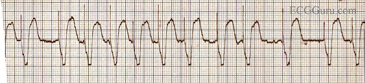
We caution students that the signs of acute M.I. (ST elevation) cannot reliably be seen in cases of wide QRS. This is because, in wide QRS situations like left bundle branch block, ventricular rhythms, or right ventricular pacing, the ST segments will elevate in leads with downward QRS complexes, and depress when the QRS is upright.  These is called discordant ST changes.


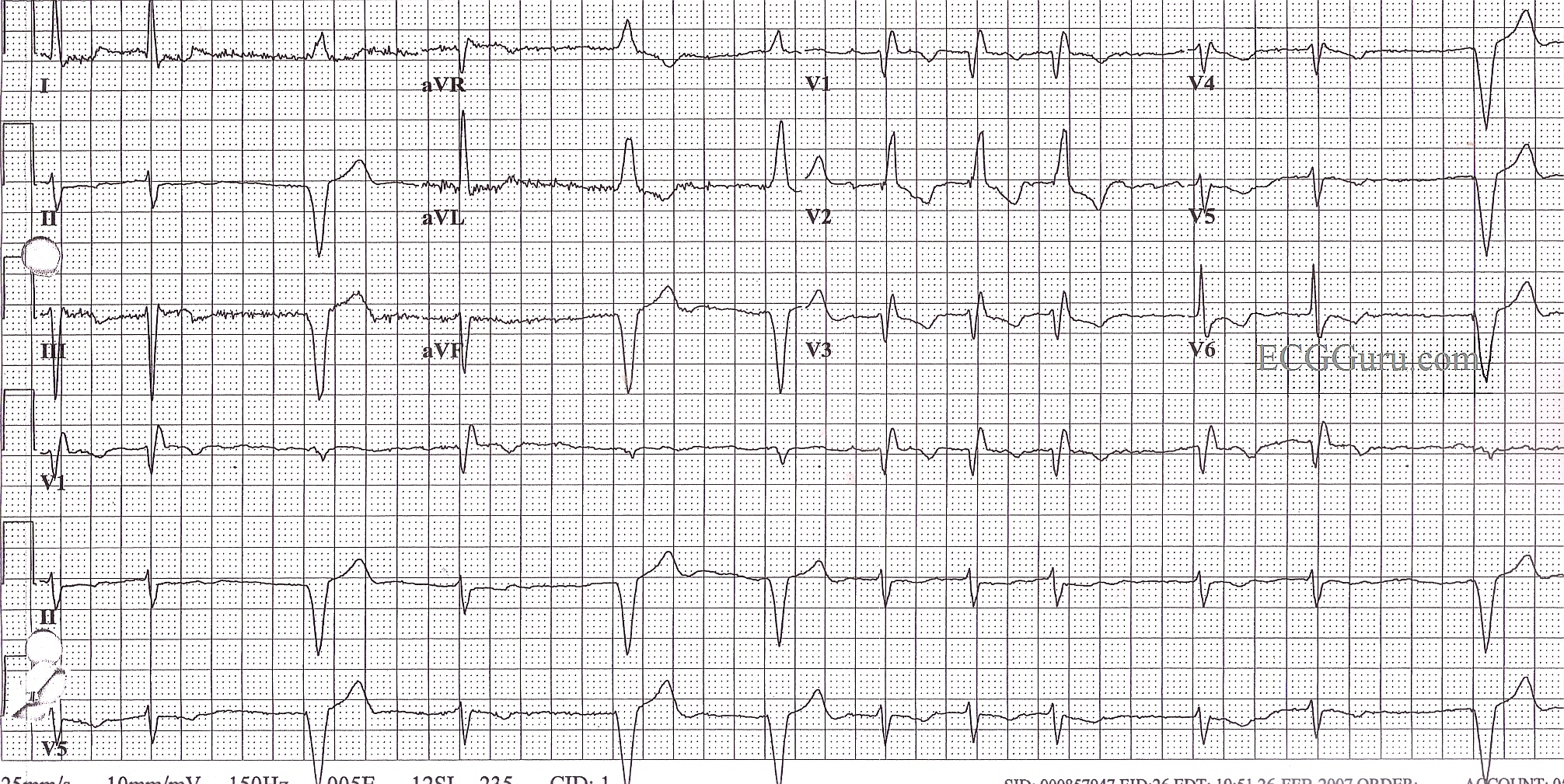
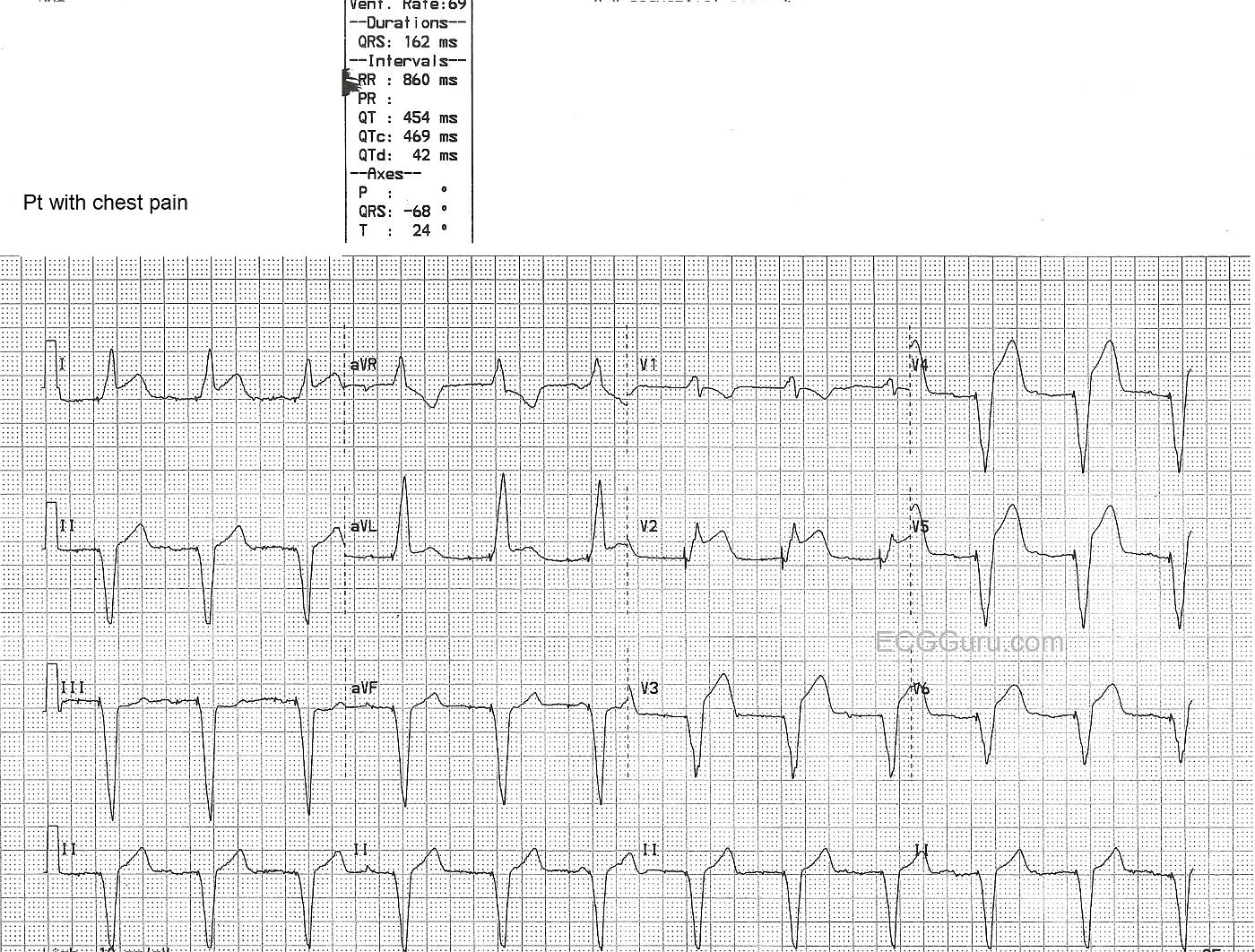
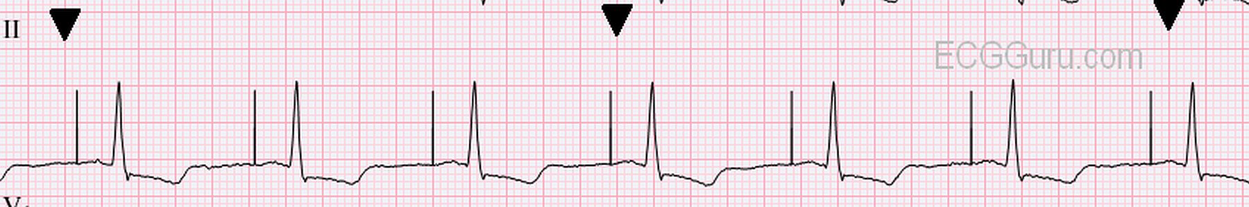
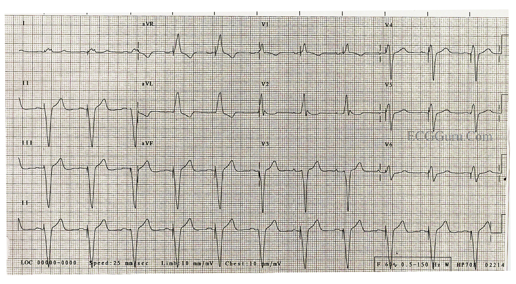
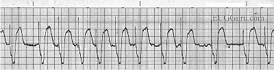
In this ECG, a man in his 60's presented with chest pain. His ECG showed AV sequential pacing, with ventricular pacing from the right ventricle. The QRS is 162 ms in duration. He has ST segment elevation in Leads I, aVL, and Leads V2 through V6.


The ST elevations are more pronounced than expected in this paced patient. But, the real clue here is the ST elevation in Leads I, aVL, and V2 - leads that should have ST depression because of their upright QRS complexes, have elevation! This patient was taken to the cath lab and the left coronary artery wass reperfused and stented. For more information about ST elevation in wide QRS complex rhythms, see this LINK.
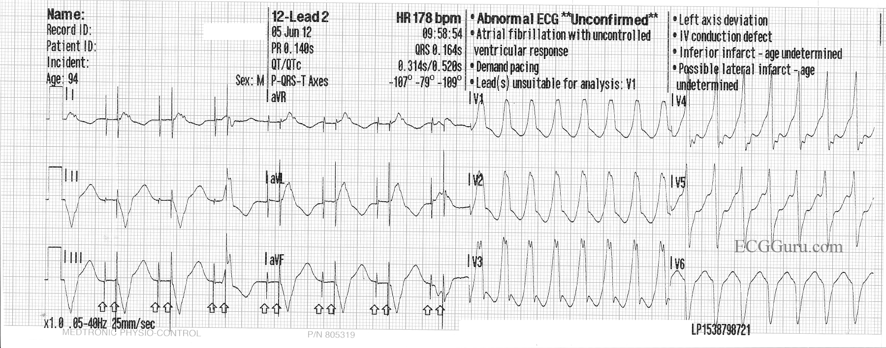
 

Pages

All our content is FREE & COPYRIGHT FREE for non-commercial use

Please be courteous and leave any watermark or author attribution on content you reproduce.