

Submitted by Dawn on Sun, 08/23/2026 - 17:13
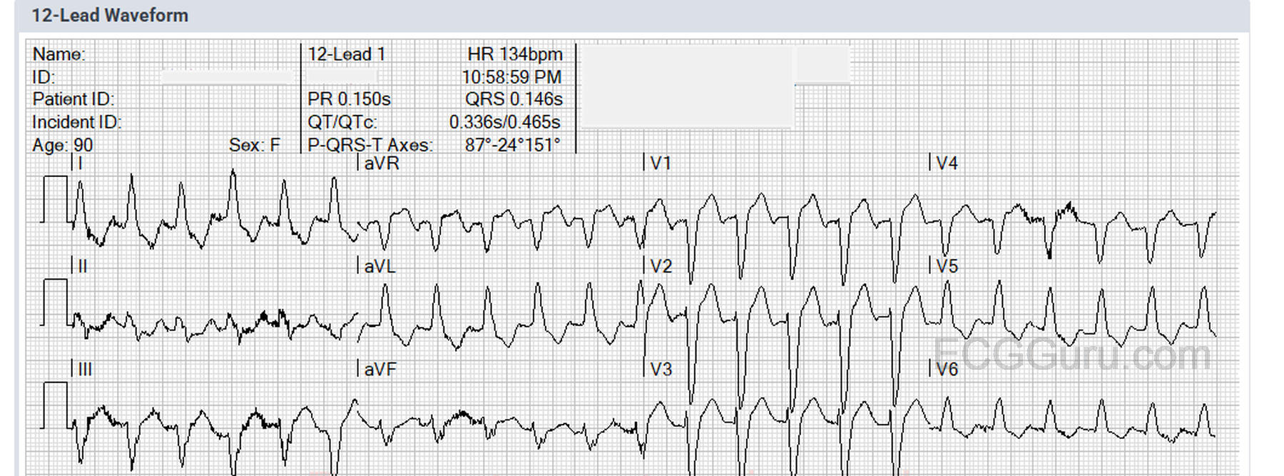
The Patient: This ECG was obtained from a 90-year-old woman who was complaining of feeling short of breath while lying in bed. Patient denied relevant medical history (respiratory or cardiac) and denied taking any medications. She also denied chest pain, nausea, or any other symptoms. She was found sitting upright in a recliner with mild subjective respiratory distress. She was completely alert and oriented, and was able to stand and transfer to the EMS stretcher. We do not have a record of her initial vital signs or lung sounds, but her initial SPO2 on room air was 80%. This improved to 100% on 15 L of O2 via NRB. She was transported to the hospital without incident.
The ECG: The ECG shows a rate of 134 bpm. The rhythm is regular. P waves are difficult to see because of the muscle tension artifact and the rate. I strongly suspect there is a P wave at the end of the T – best seen in aVR, in which you can see the negative P wave right after the T. The QRS is wide at 146 ms, and the QTc is borderline long at 465 ms, but is affected by the wide QRS being part of the measurement. There is, at this time, no simple and reliable method of evaluating QTc in the setting of wide QRS, but there are formulas available, and being evaluated.
The ECG shows a typical LEFT BUNDLE BRANCH BLOCK pattern. V1 has a negative QRS deflection, while Leads I and V6 have positive QRS complexes. There is a leftward frontal plane axis. There are discordant ST changes – the ST segments deviate in the opposite direction of the QRS complexes. Left bundle branch block is often seen in the context of heart disease, often myopathy.
Follow up: The patient was found on echocardiography to have Takotsubo cardiomyopathy. Her ejection fraction was 30%. Because of her age and frailty, a cardiac catheterization was not done. She was discharged home after three days. Video of Takotsubo cardiomyopathy: https://youtube.com/shorts/TNSdxBW1A6Q?si=eeyYZ6zkM2U7yxRD
For a more detailed look at this interesting ECG, please see Dr. Ken Grauer's comments and annotated ECG below.
Our thanks to Jessica Banks, Paramedic, for donating this tracing.
Related Terms:
Rate this content:

All our content is FREE & COPYRIGHT FREE for non-commercial use
Please be courteous and leave any watermark or author attribution on content you reproduce.
Comments
6 Pearls on this fast LBBB with Takotsubo
Today’s case features a 90-year old woman who presented for acute shortness of breath — but no chest pain. As per Dawn — her ECG shows complete LBBB. A diagnosis of Takotsubo Cardiomyopathy was made on Echo. I’ll add a few comments on her ECG and its relation to this final diagnosis.
The rhythm in today’s case is regular at a rate of ~135/minute. At first glance — P waves are not seen. Dawn drew attention to the negative deflection in lead aVR. I favor lead V1 — as I’ve found lead aVR less reliable for identifying P waves that are not evident in lead II. The RED arrows that I’ve drawn in lead V1 of my labeled FIGURE point to a small-but-distinct negative deflection before the QRS complex. Given the anatomic proximity of lead V1 to the right atrium — I’ve found a small, negative deflection that occurs before the QRS in lead V1 to provide an invaluable clue to the likely presence of sinus rhythm (sinus tachycardia) in tracings in which no other lead convinces me about the presence of P waves. (PEARL #1: Once I started looking for this subtle negative deflection in lead V1 of tachycardia tracings — I began finding it with surprising regularity.)
So the rhythm is sinus tachycardia at the exceedingly rapid rate of ~135/minute. PEARL #2: It is unusual to see sinus tachycardia in a patient with an uncomplicated acute MI. Instead, when you see sinus tachycardia in association with an acute MI — Look for SOMETHING ELSE (ie, heart failure, shock) as the cause. But especially in a patient such as today’s 90-year old woman — the presence of sinus tachycardia this fast suggests that unless this patient is in cardiogenic shock from a huge MI (and we were never told her BP), that “something else” is going on. That “something else” could be an acute pulmonary cause (this patient was markedly hypoxemic) — or as it turned out to be, it could be heart failure not from an acute coronary occlusion MI in need of PCI — but from the catecholamine surge associated with secondary heart failure in a patient with Takotsubo.
QRS morphology in today’s case is perfectly consistent with complete LBBB because: i) There is QRS widening of ≥0.12 second; — ii) Left-sided leads I, aVL, V6 — all show monophasic upright and wide QRS complexes; — and iii) Right-sided lead V1, as well as other anterior leads all manifest predominantly negative QRS complexes.
Note that S wave amplitude in lead V2 is quite deep — and that in neighboring lead V3, the S wave is cut off. Judging from the distance between the descending and ascending limbs of the S wave in lead V3 (ie, the small, vertical GREEN line I added at the bottom of the 4th QRS complex in lead V3) — it is likely that the true depth of the S wave in this lead would be much greater than what we see had the limits of the EKG paper not cut off the end of this S wave. PEARL #3: The presence of deep anterior lead S waves (as we see here) — in association with complete LBBB — should always clue you into the likelihood of significant LVH (not at all an uncommon finding in an elderly patient with LBBB).
Finally, despite the absence of chest pain — We need to assess today’s ECG for possible acute ST-T wave changes. This assessment becomes tricky — because ST depression in lateral leads, and ST elevation in anterior leads is common in association with LBBB at faster heart rates. PEARL #4: We are looking for a disproportionate amount of ST-T wave deviation, superimposed on what otherwise might be expected in a patient with LBBB and sinus tachycardia at the fast rate of 135/minute. Thus, I interpreted the amount of ST elevation in leads V2 and V3 as “NOT disproportionately elevated” considering how deep the S waves in these leads are (ie, modified-Smith-Sgarbossa Criteria are not satisfied — because the millimeter amount of J-point ST elevation is not ≥25% of the millimeter depth of the S waves in these leads). And, the amount of J-point ST depression in lateral chest leads V5,V6 is also not disproportionately excessive given height of the R waves in these leads.
However, the same is NOT necessarily true for the millimeter amount of J-point ST elevation in leads V1 and V4 — in which I’ve drawn double YELLOW arrows to indicate what appears to be the J-point in these leads. This appears to be quite close to the 25% threshold sought with modified-Smith-Sgarbossa Criteria (ie, in lead V1, I measure 3 mm of J-point ST elevation and a 12-13 millimeter deep S wave and 3/12 or 3/13 is ~25%).
Similarly, the double BLUE arrows in lead I suggest very close to this 25% threshold for the amount of J-point depression compared to the modest height of the R wave in this lead. (The 25% threshold is also quite close to being satisfied in lead aVL).
PEARL #5: In addition to seeking out the 25% threshold for disproportionate ST elevation or depression — I look for “qualitative” changes = an ST-T wave appearance that just SHOULD NOT “be there”. The one lead that I find especially suspicious of this is lead II — as there is no way the shelf-like straightened ST depression in this lead should be there.
Bottom Line Regarding assessment of ST-T wave appearance in today’s ECG — We see LBBB with ST-T wave changes of potential concern for acute occlusion in multiple leads. That said — given the extreme tachycardia, I find it hard to know if these ST-T wave changes are truly indicative of an occlusion-MI vs simply the result of a marked sinus tachycardia. But — IF consideration was being given to cardiac catheterization — then repeating the ECG within 10-15 minutes, and frequently thereafter as needed, may help to clarify whether these ST-T wave changes are or are not primarily rate-related (ie, hopefully acute treatment of this patient’s hypoxemia will result in some slowing of her heart rate, thereby allowing more objective assessment of ST-T wave appearance on subsequent ECGs).
PEARL #6: Echo sometimes SAVES the day! I see nothing on this ECG that tells me this patient had Takotsubo Cardiomyopathy. The telltale QTc prolongation that sometimes is key to suggesting Takotsubo is impossible to assess with LBBB and a tachycardia this fast. ECG findings of Takotsubo sometimes mimic the changes of acute MI. Troponins are commonly mild-to-modestly elevated with Takotsubo. But bedside Echo sometimes provides the likely diagnosis within minutes (ie, in patients with massive acute PE and RV dilatation — or in today’s case, in which Echo strongly suggested Takotsubo rather than occlusion-MI as the likely cause of the ECG abnormalities that we see). The “good news” being — that for as sick as this 90-year old woman was at the time she presented — the cardiopulmonary effects associated with Takotsubo sometimes reverse surprisingly quickly — with this 90-year old woman surviving to be discharged from the hospital after only 3 days! It’s always rewarding when cases like this have a happy ending!
P.S. For more on ECG findings and the presentation of Takotsubo Cardiomyopathy — Check out my ECG #456 (https://www.ekgblog.com/2024/10/ecg-blog-456-woman-with-chest-pain-from.... ).
Ken Grauer, MD www.kg-ekgpress.com [email protected]