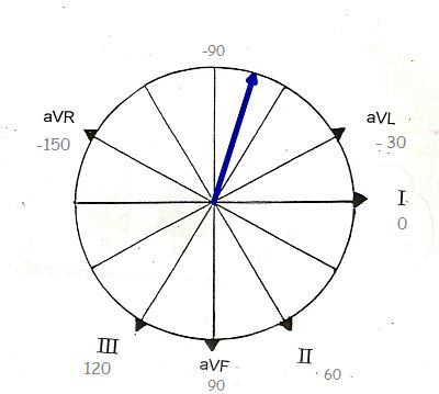
If you are teaching frontal plane axis to your students, you will need to teach them HOW to determine the axis - usually beginning with the QRS axis and then adding the P and T waves. But, you also need to teach them WHY we measure axis, to provide relevance to something that may seem challenging to beginners. There are many ECG interpretations that rely heavily or are dependent upon the determination of the axis.
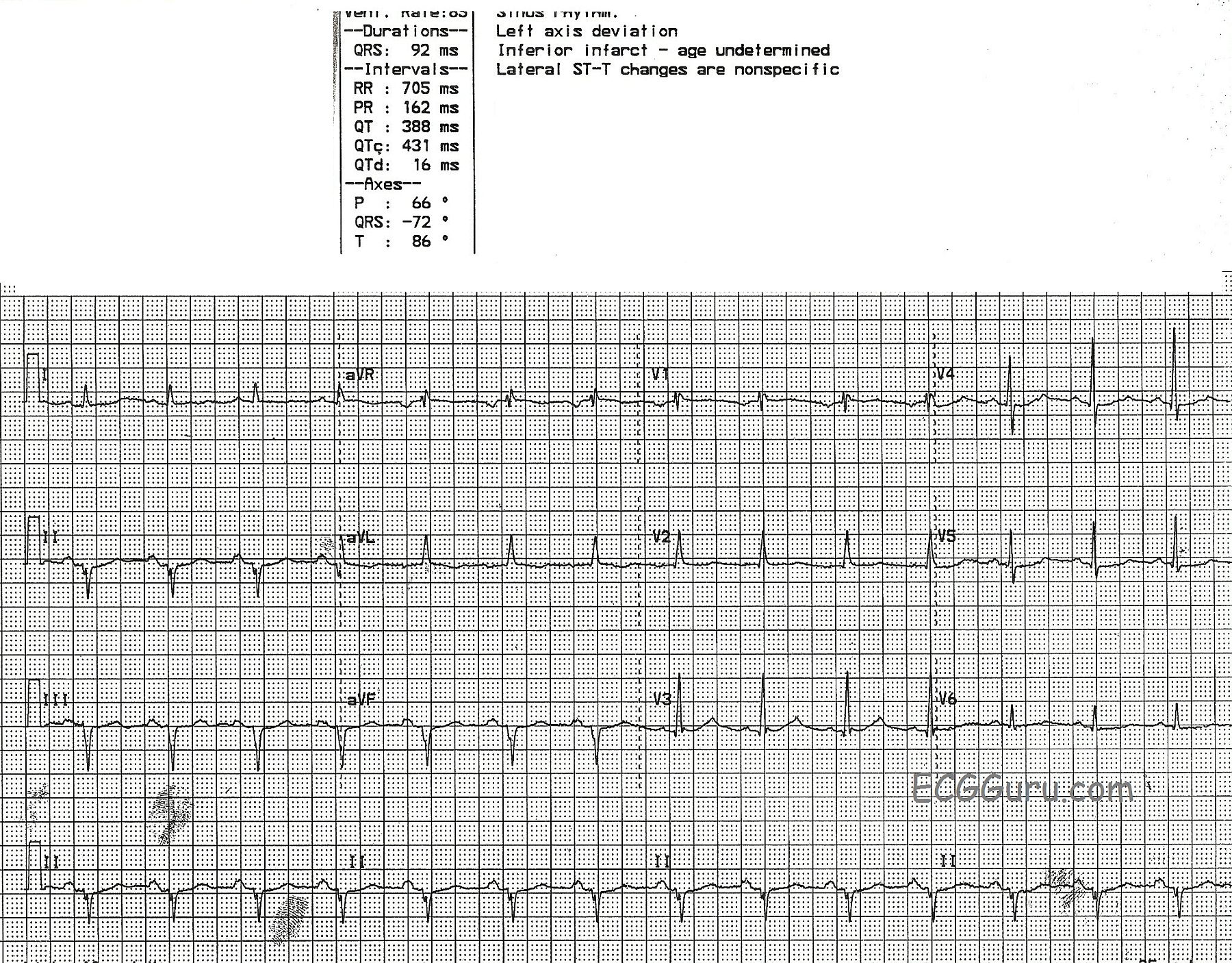
This ECG is a great example of left axis deviation. The cause is readily discernible, if your students know the ECG signs of myocardial infarction. This patient had an inferior wall M.I. in the distant past, and now has pathological Q waves in Leads II, III, and aVF. Pathological Q waves in related leads in a patient with history of M.I. are a sign of necrosis, or permanent damage, in that part of the heart. The inferior wall has lost an extensive amount of tissue, which is now electrically inactive as well as mechanically inactive. (You may also find it helpful to show students videos of ventriculograms showing normal LV function and hypokinesis of the LV due to M.I.) Because of the loss of electrical activity in the inferior wall, the "mean" electrical direction (or axis) is AWAY from the inferior wall. That is, the electricity travels AWAY from II, III, and aVF and TOWARD I and aVL.
Many of the blogs and webpages listed in our "Favorites" address the subject of axis determination. Here is one from Cardio Rhythms Online if you would like a review.