When teaching ECG, I always try to make the ECG interpretation have some practical context for the student. Why study squiggly lines, if they don't mean something to our care of our patients? Even putting a simple scenario (actual or invented) with an ECG can make it more relevant for your students. A series of ECGs taken as the patient undergoes changes, is especially helpful.
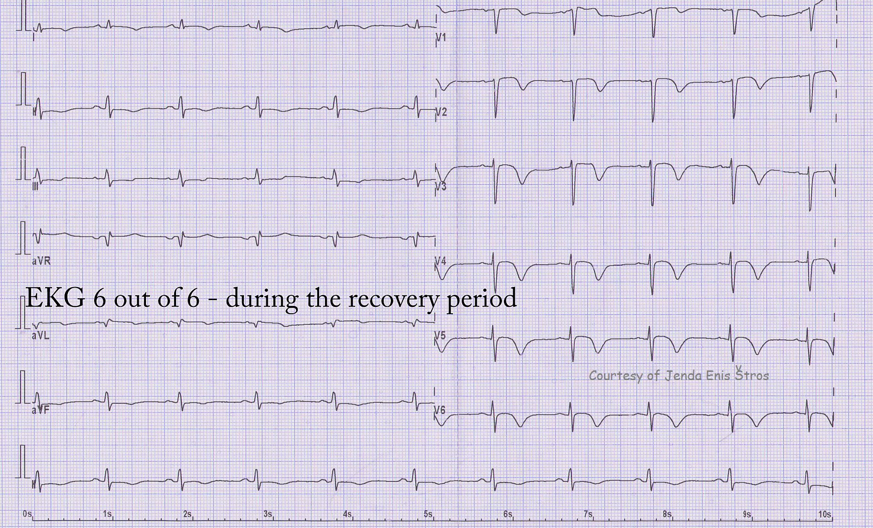
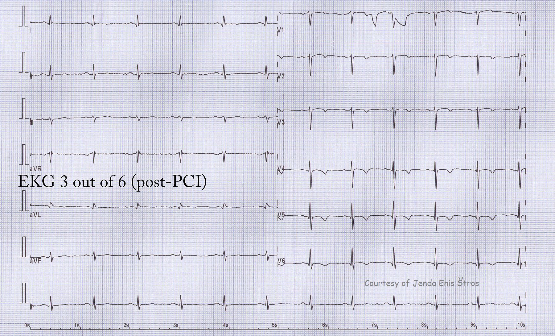
This ECG is the last in a series of 6 that were donated by Jenda Enis Štros showing the evolutionary changes of an M.I. from onset, through spontaneous reperfusion, angioplasty, re-occlusion by thrombus, and recovery. This ECG shows deep precordial T wave inversions, an expected evolutionary change after reperfusion of an occluded artery - in this case, the left anterior descending. The patient has lost some of his QRS amplitude (viable heart muscle), but has not developed pathological Q waves. Pathological Q waves would indicate full-thickness necrosis of the wall, which is usually a permanent injury.
The patient was discharged home with a 45% ejection fraction (60% is ideal), and he had akinesis of part of his anterior wall. This can be permanent or temporary, and followup studies would be needed to evaluate the ongoing health and function of the left ventricle.
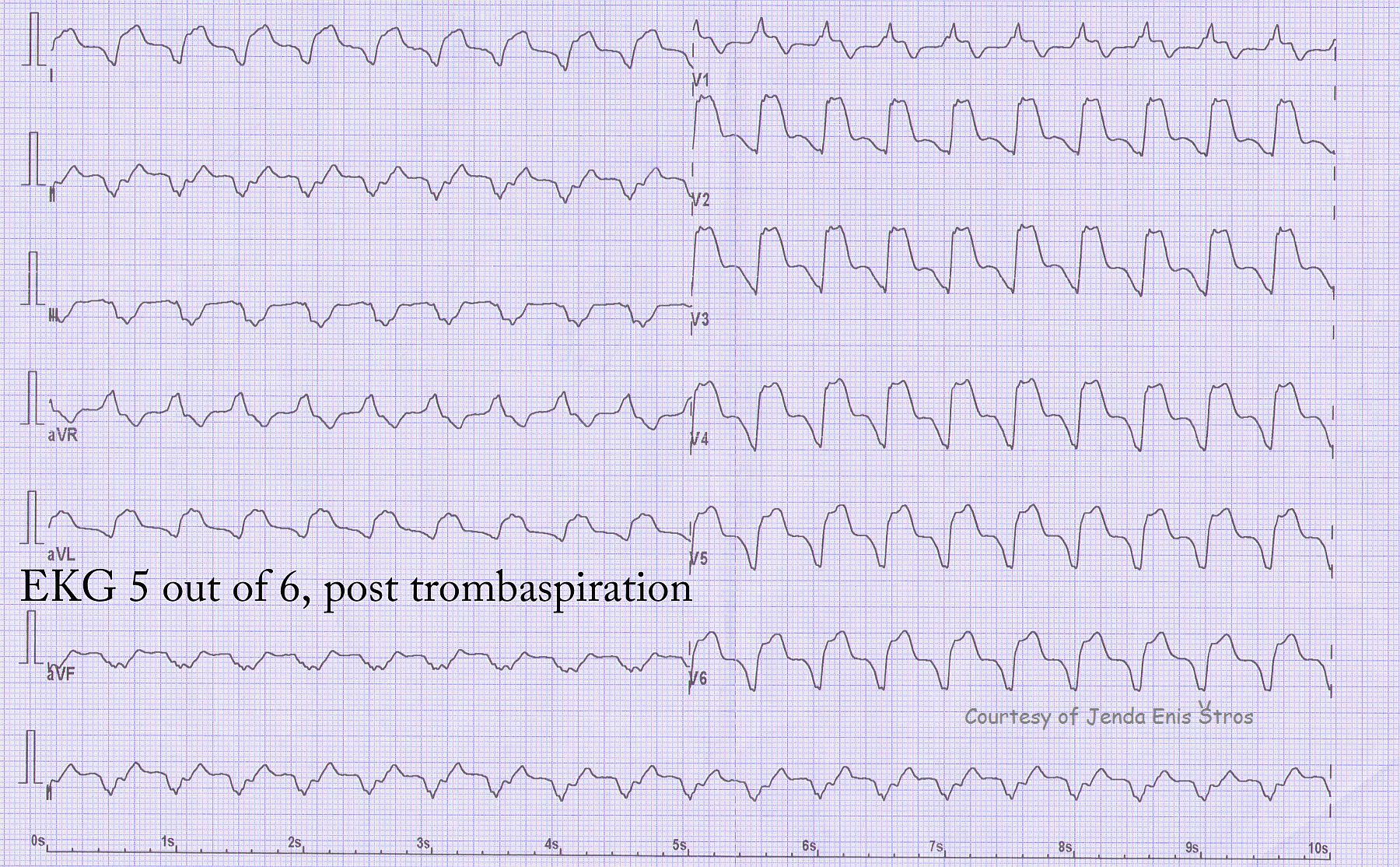
Continuing our series from the patient with acute AWMI, donated by Jenda Enis Štros, a new dysrhythmia has appeared. The patient was taken to the cath lab, and a thrombus was removed from the stent. We now see a wide-complex tachycardia. The morphology (shape and configuration) of the QRS has changed considerably from the previous ECGs, so we know this is idioventricular in origin. There are many other clues, but some major ones are: the QRS is very wide, there is no associated P wave, all the precordial leads except V1 are negative (precordial concordance), V6 is negative, and the axis is away from II, III, and aVF and toward aVR (aVR is upright).
Because the rate is about 125/min., this could be called ventricular tachycardia (V Tach) or accelerated idioventricular rhythm (AIVR). The important fact here is that this rhythm was transitory and the patient remained stable. Reperfusion dysrhythmias are not uncommon, and the patient is treated as indicated by the clinical condition.
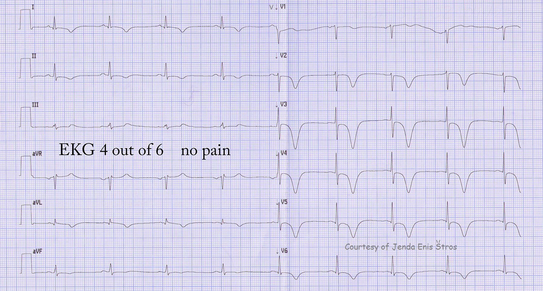
Continuing our teaching series of ECGs donated by Jenda Enis Štros, ECG 4 of 6 shows a new occurance of huge T wave inversions in the precordial leads. Since this is the area that was stented (left anterior descending artery, anterior wall of the LV), we immediately should think of re-occlusion of the artery. In a newly-placed stent, the danger is thrombosis (blood clot). The patient had no chest pain at this time.
Here are links to all six of the ECGs in this series:
This is ECG 3 in a series of 6 ECGs donated to the ECG Guru by Jenda Enis Štros. The left anterior descending artery occlusion has been confirmed in the cath lab, and angioplasty with stent placement has been performed. Post-stent, there are T wave inversions in the precordial leads (V Leads), which is an evolutionary change during the recovery phase of acute ST elevation M.I.
Here are the links to all six ECGs in this series:
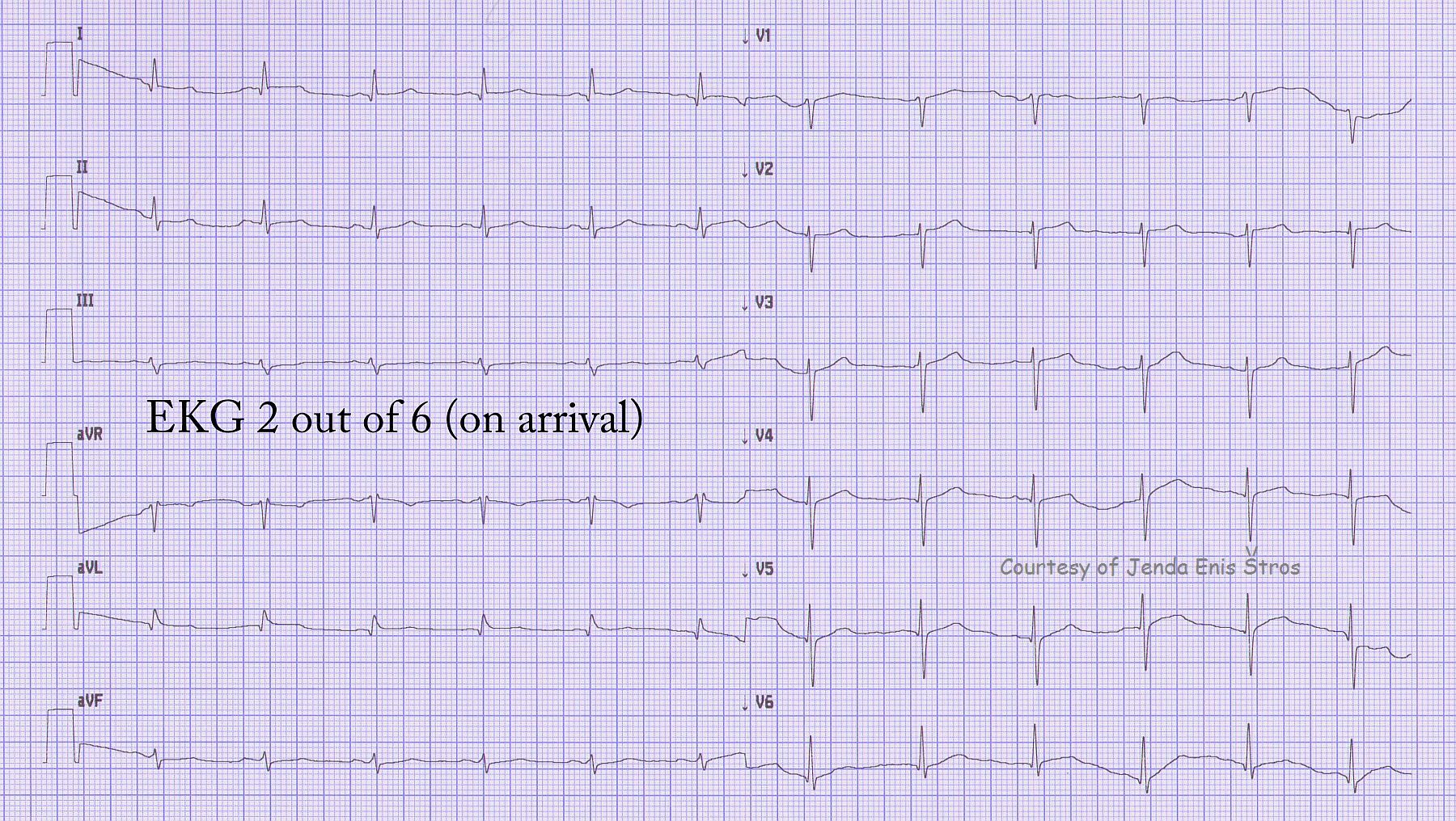
To continue the series donated by Jenda Enis Štros, ECG 2 shows spontaneous resolution of the ST elevation, coinciding with relief of the patient's chest pain. Spontaneous reperfusion can occur when the artery diameter is increased, the offending clot shifts position to allow blood flow, or other reasons. This does not mean, however, that the lesion has disappeared.
Here are the links to all six of the ECGs in this series:
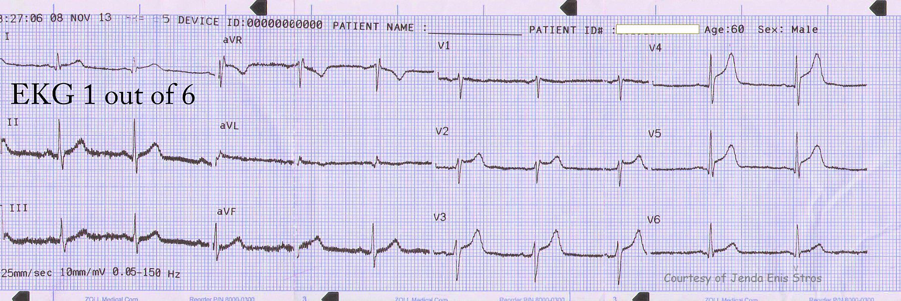
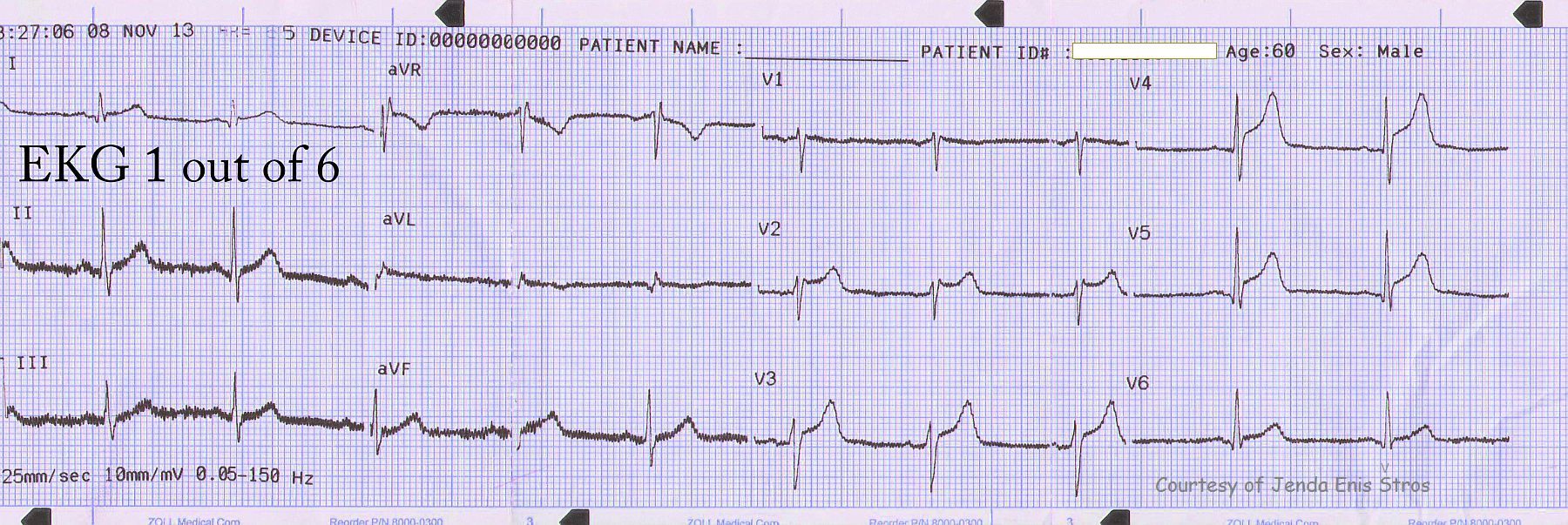
This is the first of six ECGs donated to the ECG Guru by our friend, Jenda Enis Štros.
The patient is a man in his 60's with chest pain. This is a pre-hospital ECG showing ST elevation across the anterior wall, beginning in V2. Though the upwardly-concave shape "smile" appears rather benign, the amount of j-point elevation in these five related leads - in a man with chest pain - does not favor a diagnosis of early repolarization. The diagnosis is STEMI, and the cath lab is activated.
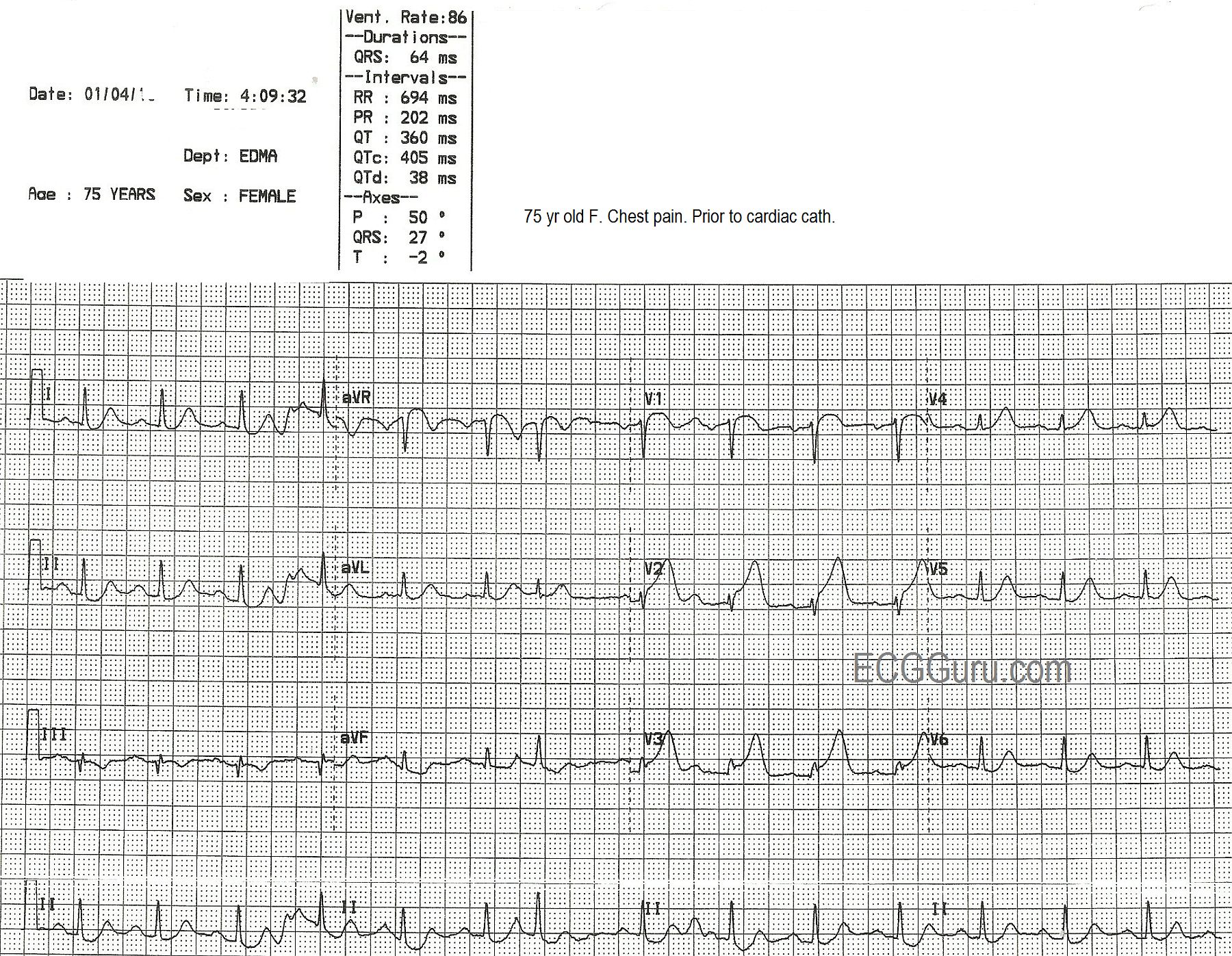
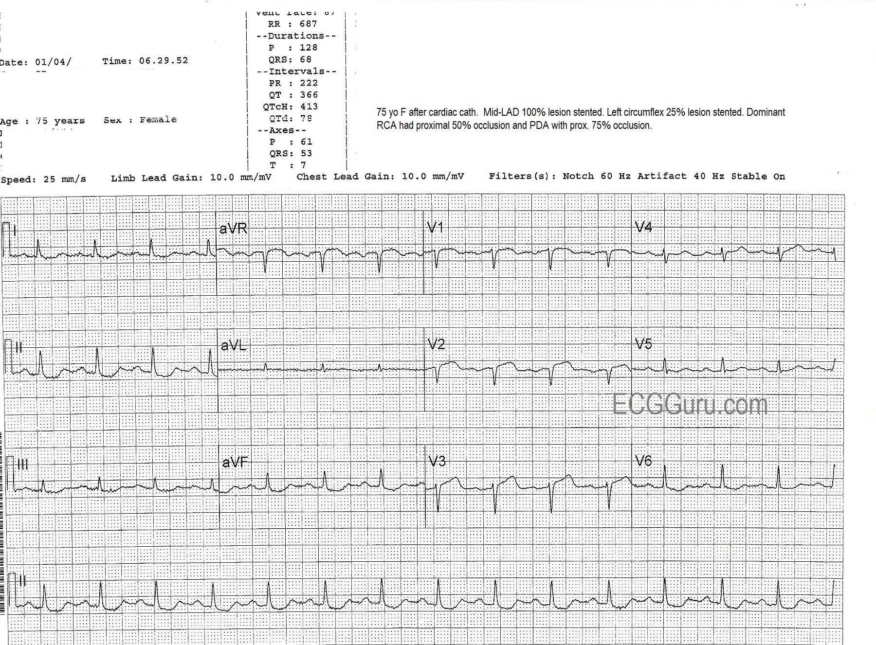
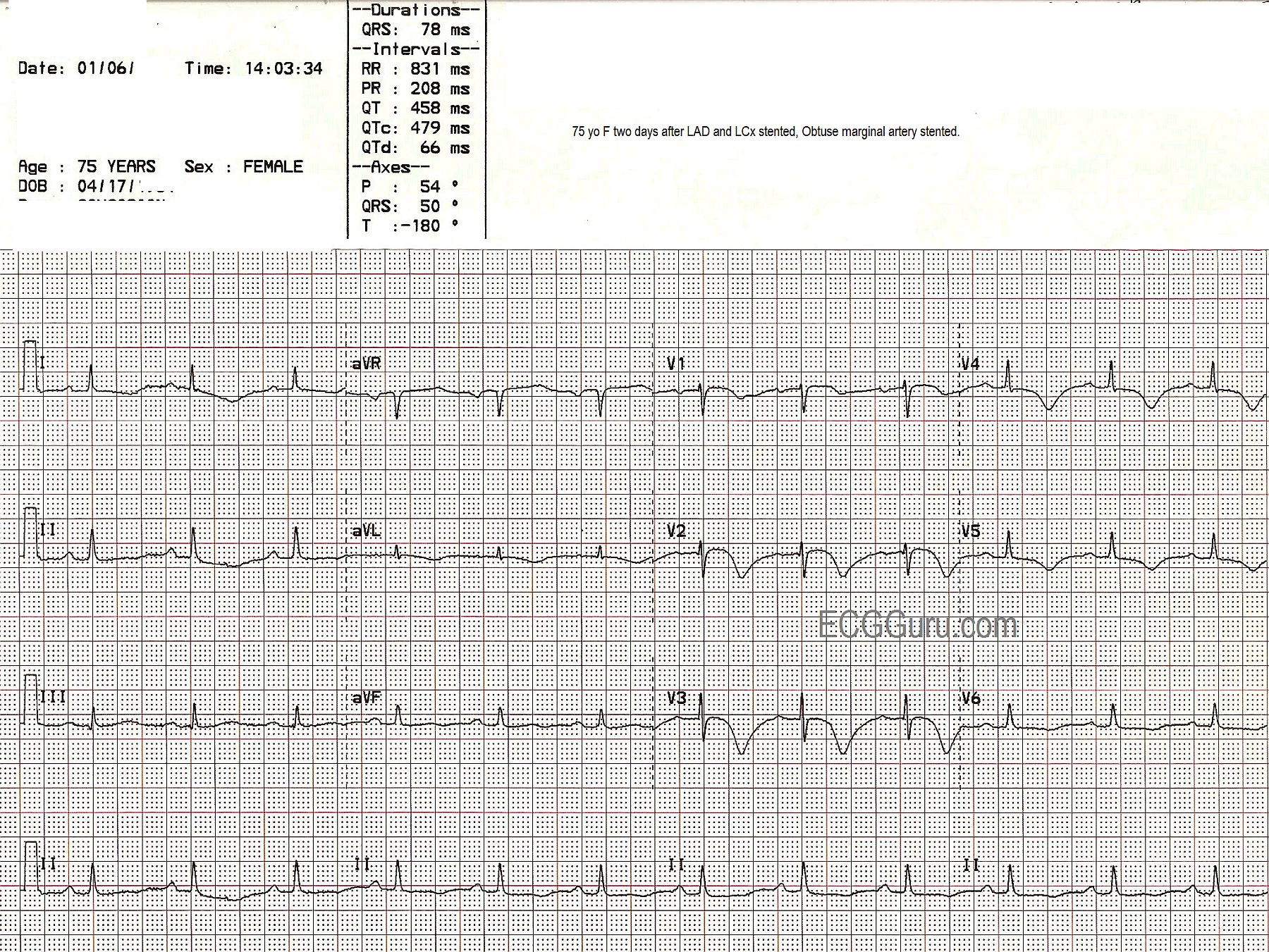
This series of three ECGs is from a 75-year-old woman who came to the Emergency Dept. with chest pain. The first ECG shows ST elevation in V1, V2, and V3, with generally low voltage in the QRS complexes. There is some coving upward of the ST segment in aVR, which can suggest a very proximal lesion of the left coronary artery (LCA). She was taken to the cath lab, where it was discovered that she had a 100% occlusion of the midportion of the anterior descending branch of the left coronary artery, which was repaired and stented. The second ECG, taken after the angioplasty, shows some Q waves in V1 and V2, with poor R wave progression in the V leads. A 25% occlusion of the obtuse marginal branch of the circumflex artery was stented two days later. The third ECG was obtained after that procedure. It shows that the Q waves have disappeared in the anterior leads (possibly due to different technicians performing the ECGs with different lead placement). It also shows marked T wave inversion in I and aVL, representing ischemia in the lateral wall, and in all the chest leads, representing ischemia in the anterior wall. The QTc is prolonged in this third ECG at 479 ms. It is not known what medications the patient was on. This patient also had a 50% proximal occlusion in the RCA and a 75% occlusion in the posterior descending artery. This is a good example of a patient with extensive coronary artery disease who finally presented for treatment when she developed ST elevation M.I.
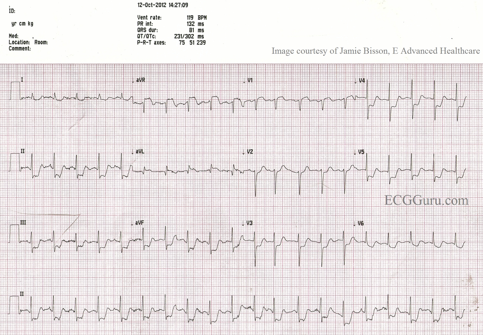
This ECG was obtained from an elderly man who was complaining of acute-onset chest pain, radiating down his left arm. He also complained of a cough, and had audible rhonchi. The machine interpretation gives several possible explanations for the widespread ST elevation noted on the ECG. The paramedics were a bit distracted by the machine's interpretation, and by the respiratory symptoms, and decided not to call a "cardiac alert" on the patient. They did, however, quickly transport him to the closest hospital, which happened to have full-service cardiac facilities. The patient was diagnosed with an acute M.I. and treated with angioplasty in the cath lab, with a good outcome. Afterward, the medics felt that they "overthought" this one, and should have given more weight to the patient's symptoms. Teach your students to evaluate their experiences with open minds and unafraid of self-criticism, so they may learn from every patient. This patient received excellent care, and the paramedics added to their "information banks", upon which they will draw for many years to come.
Although the angiogram results are not available to us, it is plausible that a proximal occlusion of the LCA, near the bifurcation of the LAD and the diagonal, could cause ST elevation in V3 through V6, with mild elevation in Lead II (which is oriented to the leftward portion of the inferior wall), and ST and T changes in the high lateral leads (I and aVL).
This ECG was provided by Jamie Bisson, of E Advanced Healthcare.
The patient, in cardiogenic shock, was resuscitated in the Emergency Department, then sent to the cath lab, where his left main coronary artery was opened and stented.
Many people with complete occlusion of the left main do not survive. When there is some diminished blood flow through the blocked area in the proximal LAD or left main, this pattern may appear. Look for ST elevation in aVR greater than or equal to 1 mm, ST elevation in aVR greater than the ST elevation in V1, and widespread ST depression.
In this ECG, aVR and V1 show ST segment elevation, with widespread ST depression. For years, aVR was virtually ignored in the literature, and considered to be only a reciprocal view of the lateral inferior wall. Now, there is convincing evidence of its usefulness in discovering proximal left coronary artery occlusion and severe triple vessel disease. ST elevation in aVR can be a reliable sign of ischemia of the basal part of the heart and the proximal IV septum.
Many people with complete occlusion of the left main do not survive. When there is diminished blood flow through the blocked area in the proximal LAD or left main, this pattern may appear. Look for ST elevation in aVR greater than or equal to 1 mm, ST elevation in aVR greater than the ST elevation in V1, and widespread ST depression.



Many people with complete occlusion of the left main do not survive. When there is some diminished blood flow through the blocked area in the proximal LAD or left main, this pattern may appear. Look for ST elevation in aVR greater than or equal to 1 mm, ST elevation in aVR greater than the ST elevation in V1, and widespread ST depression.
In this ECG, aVR and V1 show ST segment elevation, with widespread ST depression. For years, aVR was virtually ignored in the literature, and considered to be only a reciprocal view of the lateral inferior wall. Now, there is convincing evidence of its usefulness in discovering proximal left coronary artery occlusion and severe triple vessel disease. ST elevation in aVR can be a reliable sign of ischemia of the basal part of the heart and the proximal IV septum.
Many people with complete occlusion of the left main do not survive. When there is diminished blood flow through the blocked area in the proximal LAD or left main, this pattern may appear. Look for ST elevation in aVR greater than or equal to 1 mm, ST elevation in aVR greater than the ST elevation in V1, and widespread ST depression.
For complete discussions on this topic, go to Life in the Fast Lane,
Dr. Smith's ECG Blog,
JACC