Displaying 31 - 35 of 35
Dawn's picture

Inferior-posterior Wall M.I.

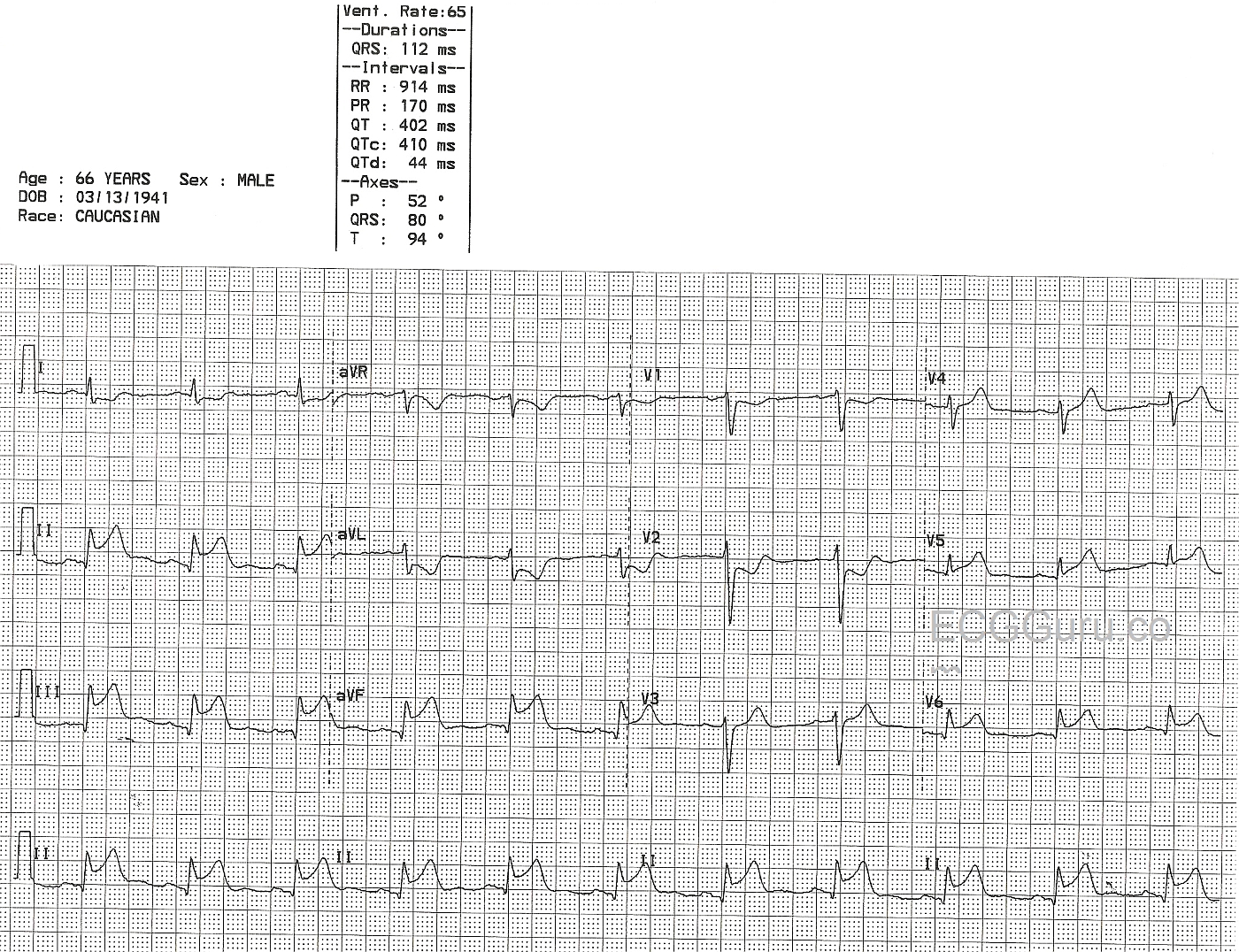
This 66-year-old man presented with a complaint of chest pain.  His ECG shows a "classic" inferior wall ST-elevation M.I. (STEMI).  The ST elevation is apparent in Leads II, III, and aVF, which are the leads that reflect the inferior wall of the left ventricle.  In addition, this ECG shows ST elevation in Leads V5 and V6, the low lateral wall.  The ST depression in V1 and V2 are reciprocal changes caused by acute injury in the posterior wall. In the majority, the RCA supplies the inferior-posterior wall, the right ventricle, the right atrium (including the SA node and the AV node), and in some, the low lateral wall.  The proximal location of this man's occlusive lesion has caused damage in all these areas.  The relatively slow rate is common in IWMI and can be caused by SA node injury, vagal stimulation, or medications.  Clinical data is not available for this patient.  

Dawn's picture

Left Bundle Branch Block With Acute Inferior Wall M.I.

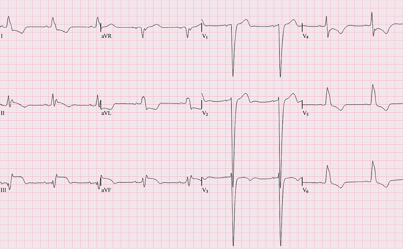
This interesting and instructive ECG was contributed by Jason Roediger, ECG Guru Extraordinaire, and one of the experts featured on our '"Ask the Experts" page. It is an excellent example of acute inferior wall M.I. with left bundle branch block. Left bundle branch block normally displays ST elevation and depression in a "negative concordance" pattern. That is, when the QRS complex is negative, we may expect ST elevation. When the QRS is positive, ST depression is seen. In this ECG, there is clearly ST elevation in Leads II, III, and aVF, and the ST segments have a distinct coved upward appearance. This shape signals to the experienced ECG interpreter that there is an acute injury.  This ECG also shows voltage criteria for left ventricular hypertrophy, seen in the right chest leads, but not the left.  

Unfortunately, the normal deviations of the ST segment seen in left bundle branch block can make diagnosis of acute M.I. difficult. For a good example of a left BBB without acute M.I., please refer to the ECG archives on this site. Often, students are taught that it is IMPOSSIBLE to see an acute M.I. in the presence of LBBB. This is not true, as this ECG clearly illustrates. See the March 4, 2012 blog post on the ECG Guru regarding this topic.

Dawn's picture

AWMI With Recent IWMI

This 88 year old woman had been sick for several days, but had not sought treatment. Her family found her nearly unresponsive and called 911. She presented to the Emergency Department as a STEMI Alert, and was in cardiogenic shock, with very poor perfusion. The ECG from the ED shows a large antero-lateral M.I., with ST elevation in V2 through V6, and also I and aVL. In addition, there are pathological Q waves, indicating necrosis, in the precordial leads, V2 through V6. The inferior wall leads, II, III, and aVF, also have pathological Q waves and abnormally shaped ST segments - no longer distinctly elevated, but coved upward.

This is a good tracing to teach students about Q waves and "old", "new", and "recent" M.I., and also about the clinical effects of hypokinesis or akinesis of the ventricles.

Unfortunately, this patient suffered a cardiac arrest in the cath lab while having her LCA reperfused with balloon angioplasty. She was resuscitated, on a ventilator and intraaortic balloon pump, and admitted to the CVICU, where she passed away within a few hours.

You will find photos from her cardiac cath in the Other Instructor Resources section, labelled as patient AW103. Click here for RCA Image, LCA Occluded Image, LCA Angioplasty Image.  Videos of her ventriculogram and left coronary artery angiogram can be found in the Resources section of this website.


Dawn's picture

Inferior - Posterior M.I.

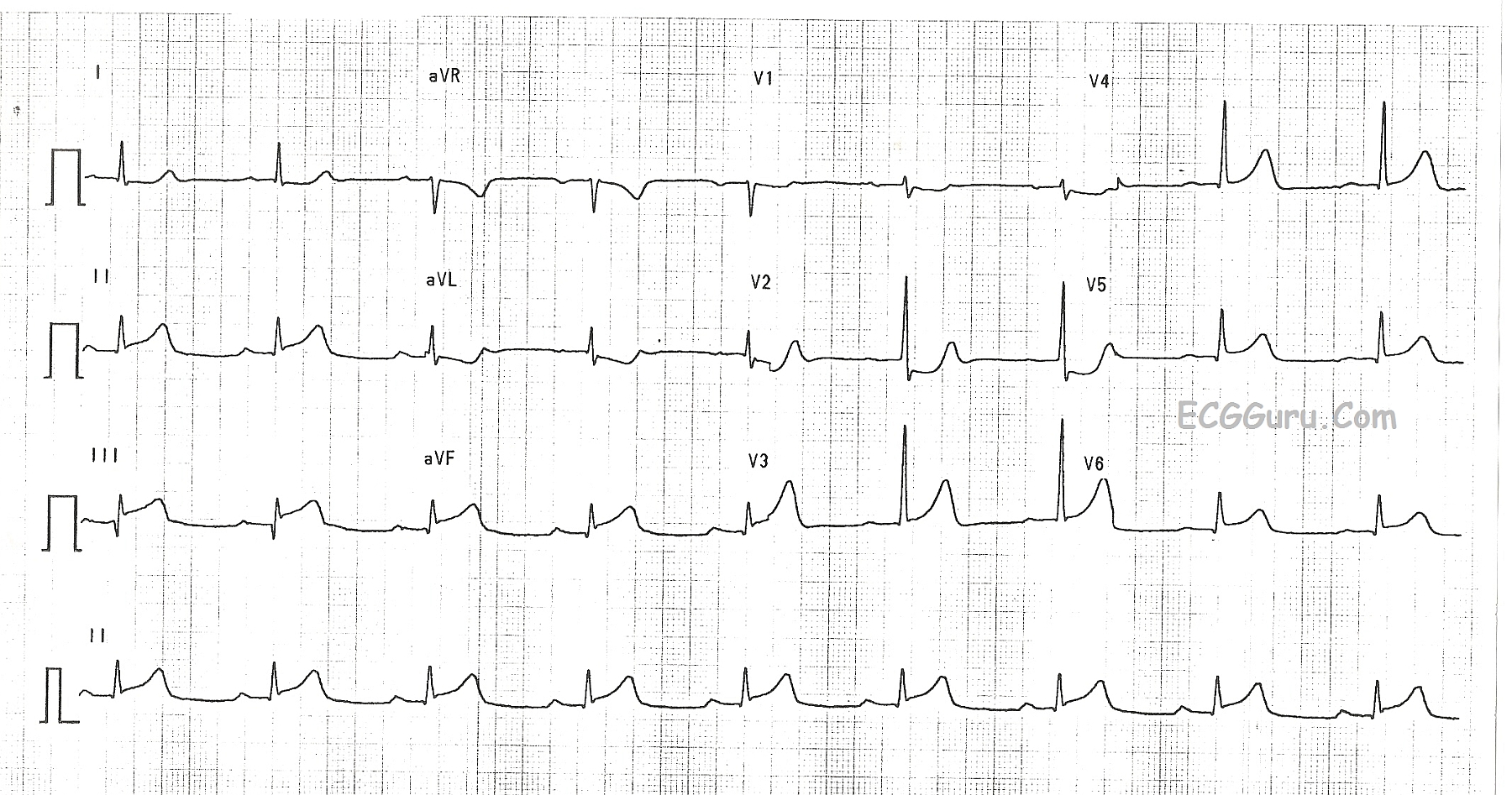
This ECG shows a classic inferior - posterior STEMI.  This M.I. was due to complete occlusion of the right coronary artery.  ST elevation apparent in Leads II, III, and aVF show the acute injury in the inferior wall, while ST depressions in V1 and V2 are reciprocal of the ST elevations in the posterior wall.  The tall R waves in Leads V1 - V3 most likely are reciprocal to pathological Q waves in the posterior wall.  Tall R waves in the right precordial leads can be caused by other cardiac conditions, such as right ventricular enlargement.  RV hypertrophy can probably be ruled out in this case because there is no right axis deviation or P pulmonale.  Because inferior wall M.I.s often extend into the posterior wall, it is the most likely cause of the tall R waves.

Dawn's picture

Inferior Wall M.I. with Right Ventricular M.I.

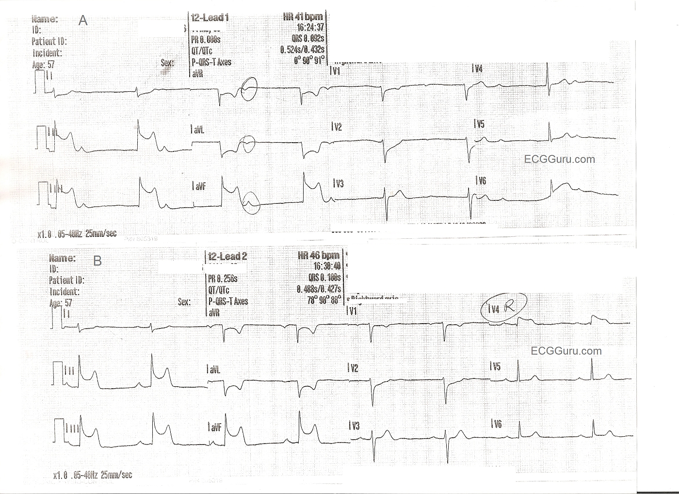
These two ECGs are from a 57 year old man with chest pain. The initial ECG shows ST elevation in Leads II, III, and aVF - inferior wall STEMI. Reciprocal changes are as expected in I and aVL. Reciprocal ST depression also seen in V1 and V2 indicate probable posterior wall involvement. Not surprising since the inferior wall is simply the lower part of the posterior wall. The first ECG also shows the patient in sinus brady with junctional escape: AV dissociation. The sinus node is often affected in IWMI that is caused by right coronary artery occlusion. The second ECG shows a slight increase in the sinus rate, and a sinus bradycardia. A V4 right lead has been performed, clearly showing ST elevation, and indicating right ventricular M.I.


FOR MORE INFORMATION ABOUT RIGHT VENTRICULAR M.I.,

Click Here

And Here

ECG Guru Resources

Pages

All our content is FREE & COPYRIGHT FREE for non-commercial use

Please be courteous and leave any watermark or author attribution on content you reproduce.