Displaying 61 - 66 of 66
Dawn's picture

ECG Basics: Atrial Fibrillation With Rapid Ventricular Response

This is a good basic rhythm strip example of atrial fibrillation with a rapid ventricular response showing the identifying characteristics of atrial fibrillation:  no P waves, an irregularly-irregular rhythm, and a "fibrillatory" baseline.  The wavy baseline will not be seen in all leads in all patients, so it is best to use the first two findings as diagnostic criteria.  Atrial fib often appears initially as a rapid rhythm, as the AV node is being bombarded by many impulses from multiple foci (pacemakers) in the atria.  Depending upon the AV node's ability to transmit these impulses,however, we could see a slow, normal, or rapid ventricular response. 

Atrial fib has very chaotic depolarization of the atrial muscle, resulting in quivering and ineffective pumping of the atria.  This loss of "atrial kick" can severely reduce ventricular filling, and can reduce cardiac output by as much as 25%.  In patients with a very rapid rate, cardiac output can be further reduced, causing CHF.  In addition, the fibrillating atria can form blood clots due to sluggish movement of blood.  These clots can embolize and cause stroke.  For these reasons, patients with atrial fib are anticoagulated and sometimes the atrial fib is stopped by medical, surgical, or electrical therapy.  Recurrence of atrial fib is common after treatment, and for some patients, control of the ventricular rate and anticoagulation become the preferred treatment.

Dawn's picture

ECG BASICS: Second-degree AV Block, Type II

Today's basic rhythm strip illustrates second-degree AV block, Type II.  Even though there is fine baseline artifact present, it is easy to measure the P-to-P interval, and your students will be able to see that every third P wave falls in the T wave.  The PR intervals are constant and the atrial rate is about 110/min.  The ventricular rate results from a 3:1 conduction ratio, and is less than 30/min.  For your students who have learned about bundle branch block, this strip shows a right bundle branch block, which is very common in second-degree Type II blocks, as they usually represent "intermittent tri-fascicular block" - that is, two of the three fascicles in the bundle branches are blocked, and one is intermittently blocked.  Other combinations of complete block and intermittent block are possible, resulting in intermittent failure of conduction.  This strip can start a lively classroom discussion about treatment of bradycardias.  See comments below for discussion of terminology, second-degree AVB and high-grade AVB.  This strip can be used as a good example of high-grade AVB. 

Dawn's picture

ECG Basics: Supraventricular Tachycardia

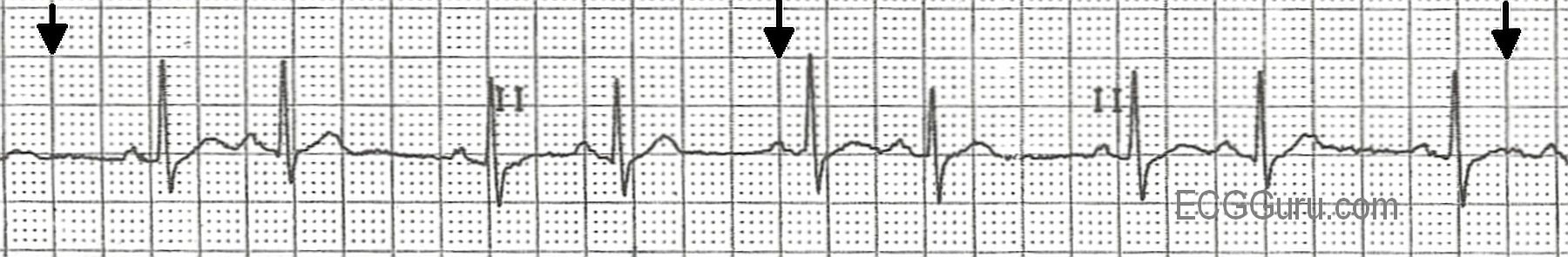
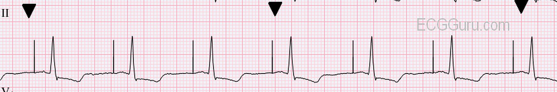
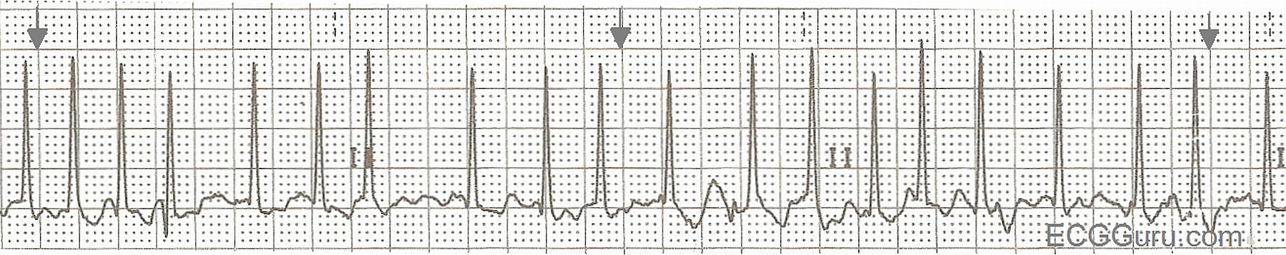
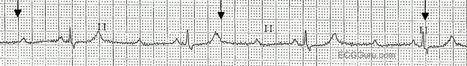
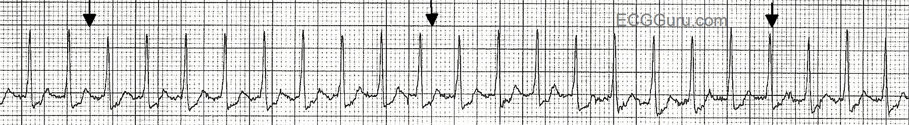
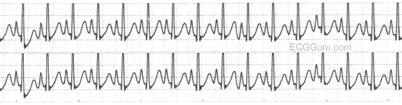
This strip is from a patient who experienced a sudden onset of palpitations and rapid pulse while at rest.  It shows a narrow-complex tachycardia, specifically a paroxysmal supraventricular tachycardia.  The subject of supraventricular tachycardias is a fascinating one, and is covered extensively throughout this website.  The mechanisms of SVT are many, and can be complex for the beginning student to understand.  Search the search terms on the left side of the page for entries from ASK the EXPERT and JASON's BLOG for more advanced information about SVT.

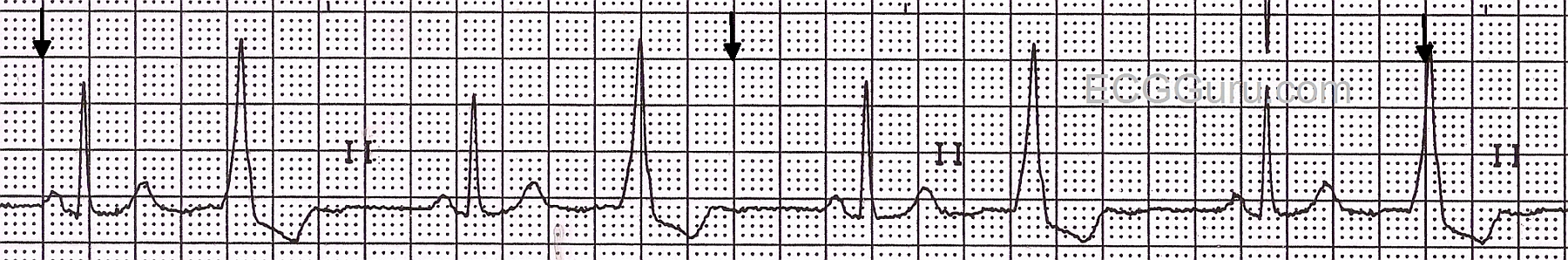
For the beginner, it is important to teach the difference between sinus tachycardia and "supraventricular tachycardia".  Of course, sinus tachycardia IS supraventricular - but current convention has us using the term "SVT" for atrial or junctional tachycardias, and especially for reentrant tachycardias.  Beginner students should understand the function of the sinus node, and it's ability to control the heart rate, based on direction given by the nervous system.  The sinus node increases and decreases the rate incrementally, or more gradually than the onset and offset of a reentrant tachycardia.  The appearance of a sudden onset of regular tachycardia following a PAC, producing a rhythm with a distinctly faster rate than the original sinus rhythm, is a sure sign of SVT.  When the onset or offset are caught on the rhythm strip, our job is SO much easier!

The heart rate helps with the diagnosis.  SVTs tend to be faster than sinus tachycardias.  SVTs tend to be faster than 150/min, while sinus rhythms TEND to be slower than 150.  And patients with sinus tachycardia usually have a readily determined reason for the tachycardia, such as fever, pain, fear, hypovolemia, hypoxia, or exertion.  So, a patient on a treadmill for an exercise stress test might very well have a heart rate over 150 / min.

When your students master the understanding of the different behaviors of the sinus node and the reentrant rhythms, and how important patient presentation is to the diagnosis, you will want to add atrial flutter and atrial fibrillation.  When these are mastered, be sure to remind your students that atrial flutter can conduct 2:1, and will mimic sinus tach.  A good rule of thumb is: Under 150/min.:  look for sinus tach first.  Around 150 / min.: look for atrial flutter with 2:1 conduction.  Over 150/min.: suspect PSVT.   


Dawn's picture

ECG BASICS: Sinus Rhythm With Ventricular Bigeminy

Nice, clear example of ventricular bigeminy with an underlying sinus rhythm.  We do not know from this strip if the sinus rhythm is a bradycardia at a rate of about 42 per minute, or if the underlying sinus rhythm is actually at a rate of 85 per minute, with every other sinus beat inhibited by the occurance of a PVC.  In the first possibility, the ventricular beats would be considered "escape" beats, positively contributing to the patient's heart rate.  In the second instance, the rather late-occurring PVCs would cause the heart to be refractory, preventing the sinus P wave from conducting it's impulse to the ventricles.   Sometimes, we can see signs of the sinus P wave "hiding" in the PVC, but in this case, if P waves exist, they fall almost exactly in the middle of the ventricular beats' QRS complex, making them invisible.  A good strategy would be to watch the strip continuously for some time, hoping to catch the conduction of two sinus beats in a row, solving the dilemma.

Dawn's picture

ECG BASICS: Sinus Tachycardia in a Toddler

Here is a nice example of sinus tachycardia taken from a 2-year-old in the post-anesthesia care unit after a short GI endoscopic procedure.  Would you call this NSR, since it is from a child?  The pre-op heart rate in this child was 120/min.

For your more advanced students, remind them that, in adults especially, a heart rate close to 150/min. should cause them to examine the ECG in several leads, looking for the presence of atrial flutter with 2:1 conduction.  Another important teaching point, most ADULTS with sinus tach at 150/min. would  manifest an obvious reason for the rapid heart rate (dehydration, pain, anxiety, shock, etc.)  Challenge your basic students to come up with as many causes for sinus tach as they can.

Dawn's picture

ECG BASICS: Sinus Bradycardia With First-degree AV Block

TODAY, we are starting a new feature on the ECG GURU.  ECG BASICS will provide rhythm strips and 12-leads for your beginner or refresher students.  It can be discouraging to the entry-level student to see only intermediate or advanced material and not understand it.  We must remember to start at the most elementary concepts, and then build on them, just as we do with any other subject.  Even more advanced students sometimes benefit from a return to the "basics".  In this weekly feature, you will find downloadable content that is, like all ECG Guru content, FREE for use in an educational context.  Please let us know in the "Comments" section below what ECGs, rhythm strips, or illustrations you would like to see featured in this new area.


Today's strip:  Sinus bradycardia with first-degree AV block.  The rate is in the 30's and slowing, and the PR interval is .26 seconds.

Pages

All our content is FREE & COPYRIGHT FREE for non-commercial use

Please be courteous and leave any watermark or author attribution on content you reproduce.