Displaying 11 - 20 of 20
Dawn's picture

Sinus Tachycardia in a Child

A six-year-old girl was found with her two younger siblings and her mother, unconscious, in a room filled with carbon monoxide.  The mother had been using a charcoal grill inside the house.  She managed to call 911 before losing consciousness, and the fire rescue paramedics broke into their house, saving them.  This six-year-old required treatment in the pediatric intensive care unit, but recovered completely.  The lesson for our students is to judge rate in the setting in which it is found.  The heart rate here is 136/min.  Normal for a child of six is usually between 80 and 100 at rest.  This child is severely hypoxic, and she has sinus tachycardia.  It would be a mistake to assume her rate is normal because "all children have fast heart rates".  Also, children with sinus tachycardia can be so fast, they appear to have PSVT.  The onset and offset can be excellent clues to the origin of the rhythm.  Sinus rhythms can be expected to speed up and slow down gradually, unlike PSVTs, which have sudden onset and offset.  The most important consideration is that sinus tachycardia usually has an APPARENT CAUSE:  exercise, anxiety, hypoxia, hypovolemia, fear ,,,, the list is a long one.  Once it is determined that the patient has sinus tachycardia, efforts usually focus on the elimation of the cause.


We welcome any comments on this ECG, perhaps taking the discussion to a more advanced level.

Dawn's picture

ECG BASICS: Sinus Tachycardia in a Toddler

Here is a nice example of sinus tachycardia taken from a 2-year-old in the post-anesthesia care unit after a short GI endoscopic procedure.  Would you call this NSR, since it is from a child?  The pre-op heart rate in this child was 120/min.

For your more advanced students, remind them that, in adults especially, a heart rate close to 150/min. should cause them to examine the ECG in several leads, looking for the presence of atrial flutter with 2:1 conduction.  Another important teaching point, most ADULTS with sinus tach at 150/min. would  manifest an obvious reason for the rapid heart rate (dehydration, pain, anxiety, shock, etc.)  Challenge your basic students to come up with as many causes for sinus tach as they can.

jer5150's picture

Jason's Blog: ECG Challenge of the Week for March 24th - 31st.

Patient's clinical data:  47-year-old black man.  Another serial ECG (seen below) showed a supraventricular tachycardia (SVT) at a rate of 164/min that was diagnosed by an Electrophysiologist as atrioventricular nodal reentrant tachycardia (AVNRT).  How does knowing the mechanism of AVNRT help you in determining the nature of the mechanism in this week's ECG.  

jer5150's picture

Jason's Blog: ECG Challenge of the Week for Feb. 24th - March 3rd.

Patient's clinical data:  55-year-old white man admitted to the surgical intensive care unit (SICU). 

Part of the computer's interpretation was:
Sinus tachycardia 1st degree AV block Occasional
Premature supraventricular complexes

DO YOU AGREE WITH THE COMPUTER?

Dawn's picture

Sinus Tach vs SVT In An Inebriated Patient

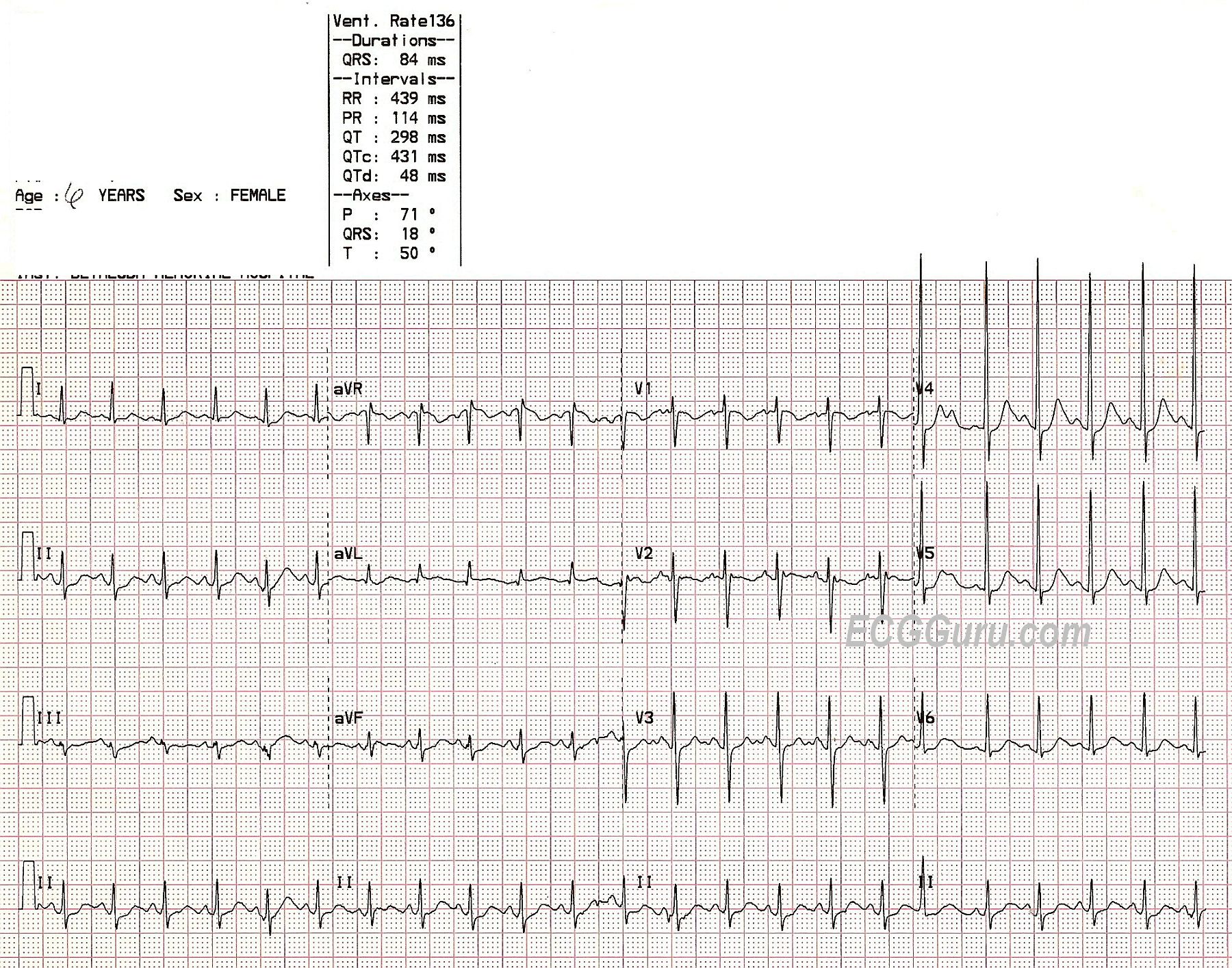
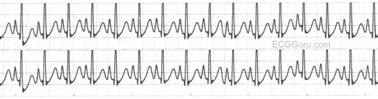
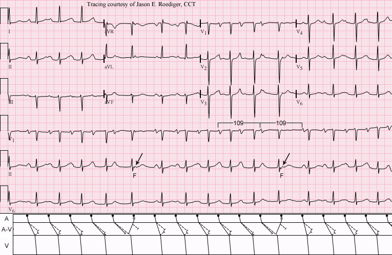
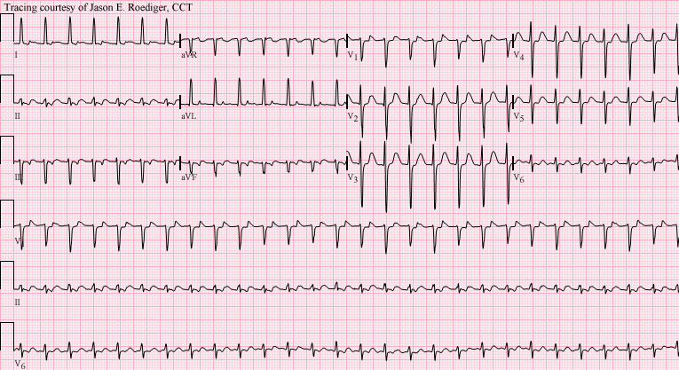
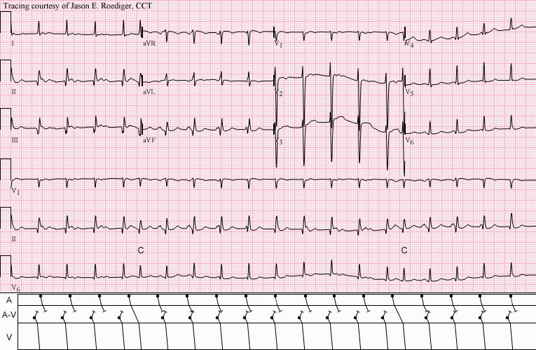
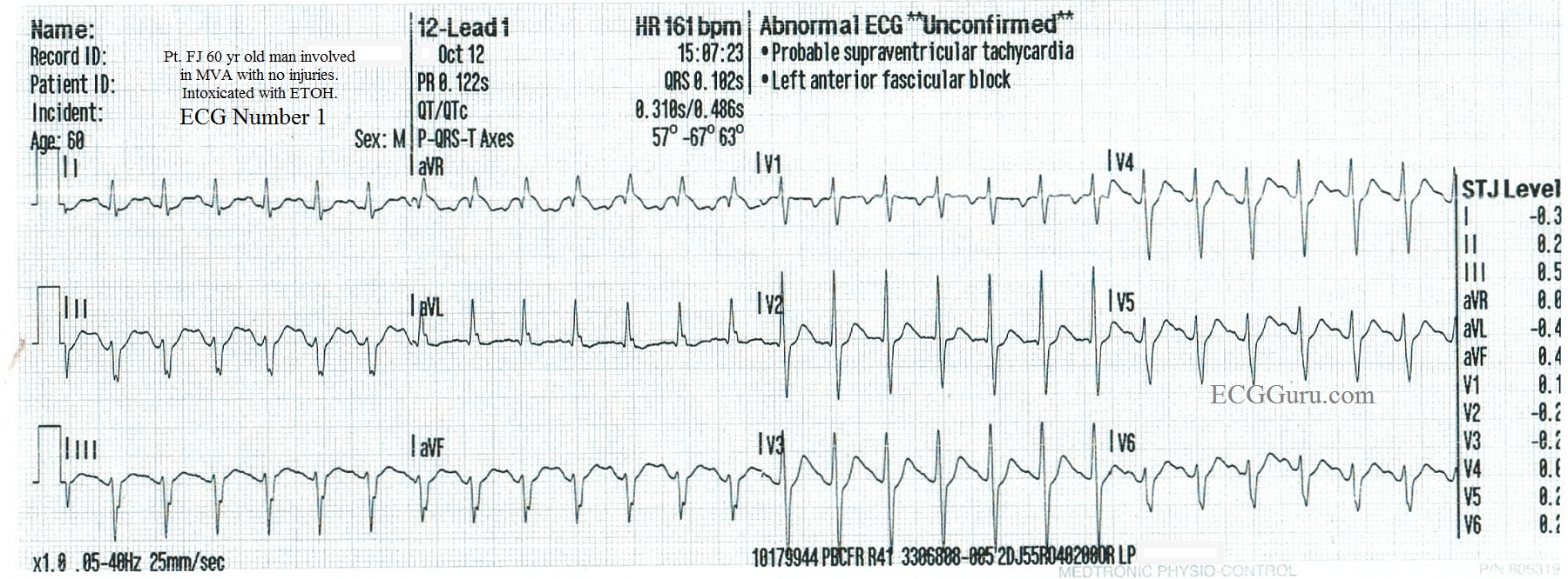
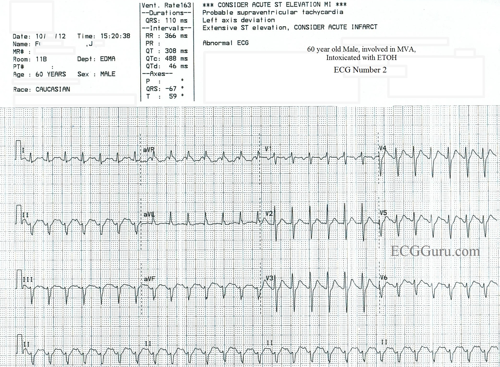
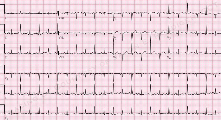
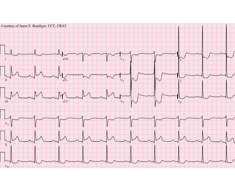
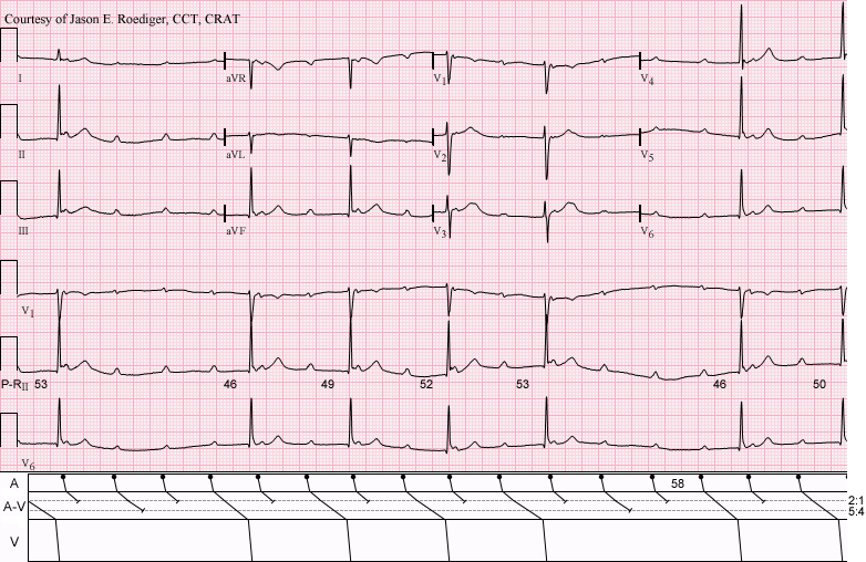
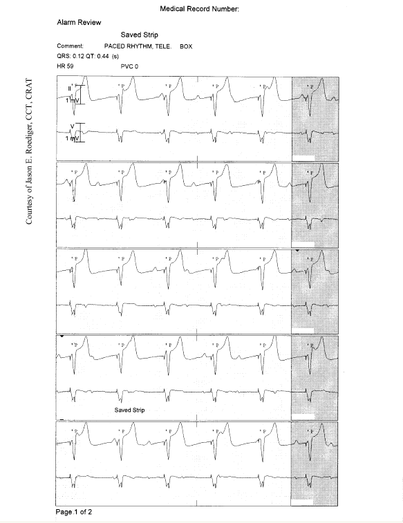
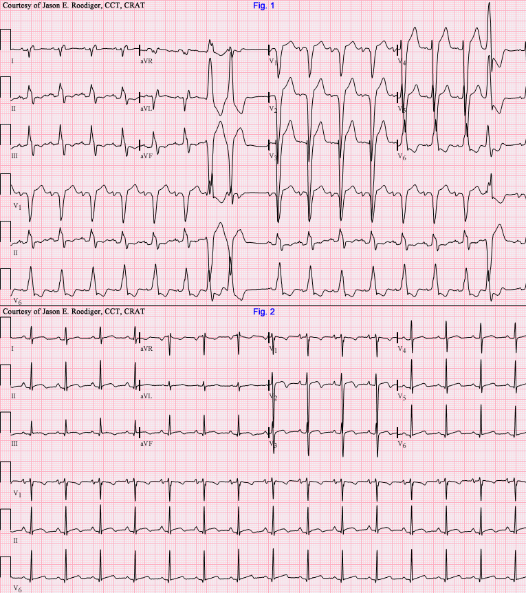
This series of ECGs was obtained from a 60-year-old man who was involved in a one-car accident.  He sustained no injuries, but his blood alcohol level was far above the legal limit for intoxication at over 300 mmol/L.  ECG No. 1 shows the ECG obtained by paramedics in the field, which they incorrectly interpreted to be atrial fibrillation.  No medication was given.  The ER physician obtained ECG No. 2, and considered sinus tachycardia as the diagnosis, but also, because of the fast rate and the fact that the rate had not changed for at least 15 minutes, he considered SVT or atrial flutter with 2:1 conduction.  The ERP administered diltiazem (Cardizem) to the patient, which resulted in ECG No. 3.  The transition to the slower rate was not captured on rhythm strips, but the nurse's notes showed a gradual change over 15 minutes from a rate of 160 to 105/min.  

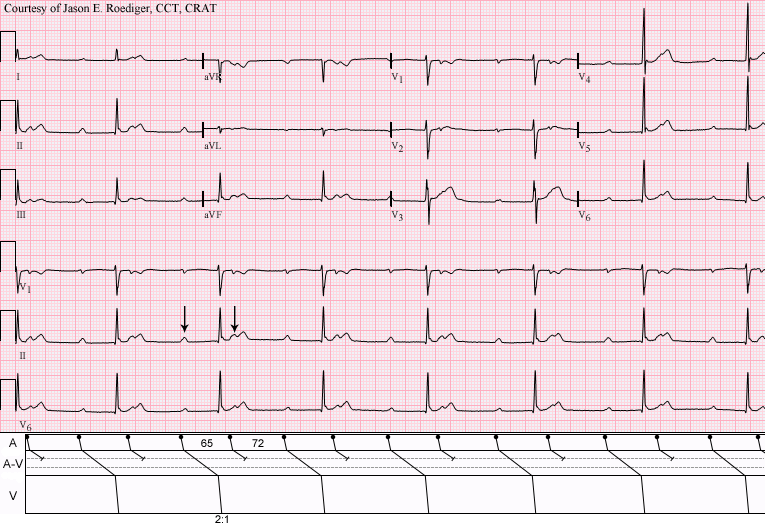
Usually, on the Instructors' Collection ECGs, we like to give the "answer".  In this case, however, there will undoubtedly be some discussion regarding what went on.  This discussion can be useful if you are teaching intermediate to advanced students.  Questions to consider:  1) Is the fast rhythm an SVT and, if so, which one?  2) Is it sinus tachycardia and, if so, what are the effects of the car accident and the alcohol?  3) Is the left anterior fascicular block relevant? (Criteria are left axis deviation, slightly widened QRS complex at 110 ms, no other obvious reason for the axis deviation). 4) Is the ST elevation in the inferior wall during the tachycardia a sign of acute M.I.?  The patient was lost to followup, so it is not known whether the ST changes were investigated.  Note the flat ST segment and inverted T waves in V1 during the tachycardia that resolve when the rate decreases. 

Please log in to register your comments on this interesting series.

jer5150's picture

Jason's Blog: ECG Challenge of the Week for Sept. 30th - Oct. 7th.


This is another one of those instances where I fortuituously viewed this ECG the day it was performed in our triage department several years ago. I immediately knew what was happening here but apparently nobody in the emergency department saw the obvious clues.

jer5150's picture

** *** ACUTE MI *** ** (but what else?)

INTERPRETATION:
(1.)  Sinus tachycardia (rate about 114/min) with . . .

(2.)  . . . acute inferoposterior infarction complicated by . . .

jer5150's picture

LYME DISEASE CAUSING AV BLOCK

We are now in the month of August.

Dawn's picture

Wide Complex Tachycardia: Left Bundle Branch Block

Today, we are revisiting an ECG from the archives, with a NEW comment from Dr Ken Grauer. This ECG is worth a second look because it is a very good example of the left bundle branch block pattern. It's also a good ECG to use when discussing treatment of wide-QRS tachycardias.

This ECG demonstrates a wide complex tachycardia with classic signs of LBBB: wide QRS, QRS negative in V1 and positive in V6 and Lead I, and supraventricular rhythm. P waves are difficult, if not impossible, to discern for sure. The machine does give a P wave axis and PR interval.  When evaluating a wide complex tachycardia, the patient's hemodynamic stability will initially determine treatment.  All WCTs should be treated as V TACH until proven otherwise.  The presence of a typical LBBB pattern makes LBBB very likely, but is not a sure thing.  This patient was confirmed to have LBBB when the rate slowed, the P waves became visible, and the QRS complexes did not change.

Pages

All our content is FREE & COPYRIGHT FREE for non-commercial use

Please be courteous and leave any watermark or author attribution on content you reproduce.