Dr A Röschl's picture

SMART WATCH ECG

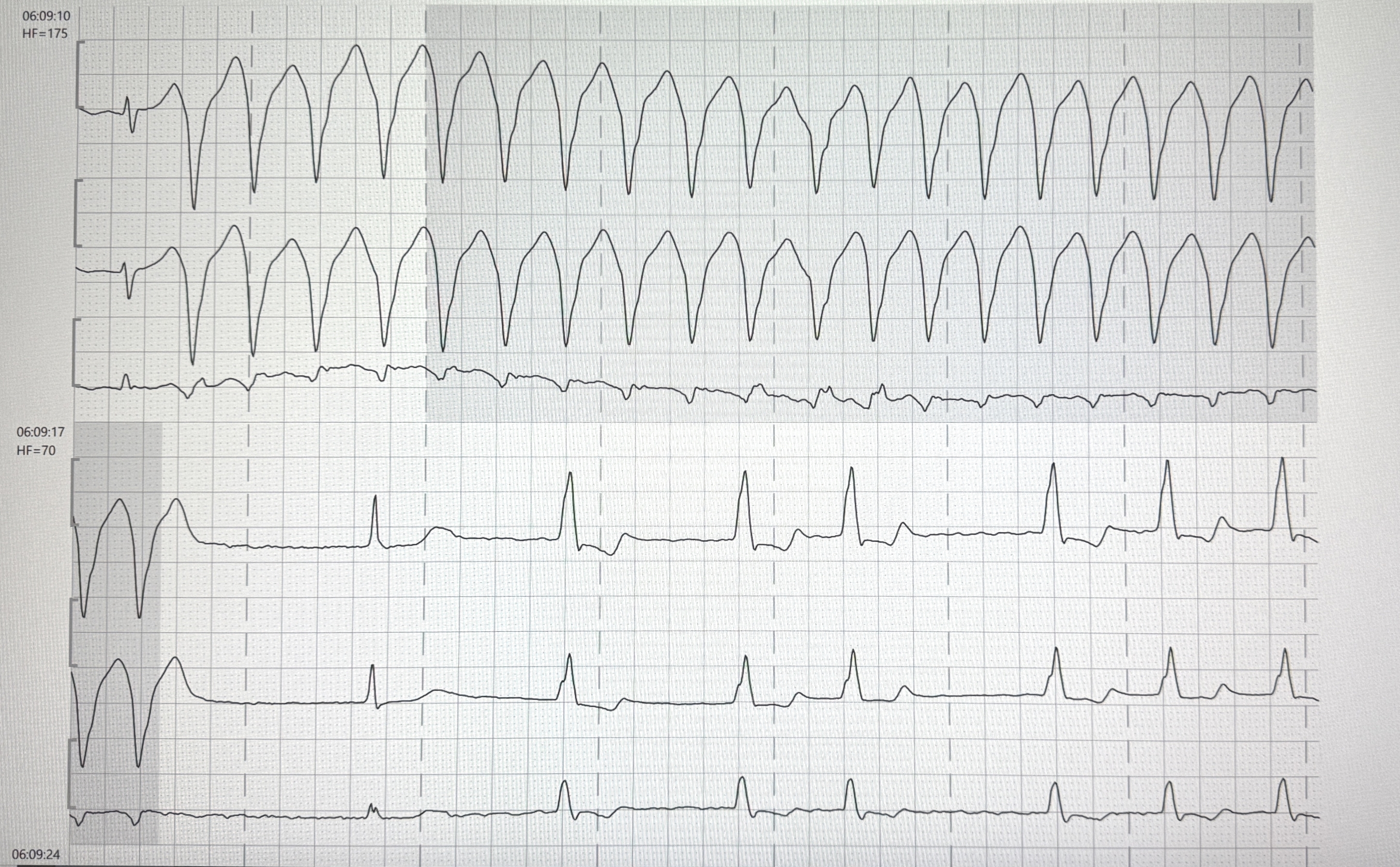
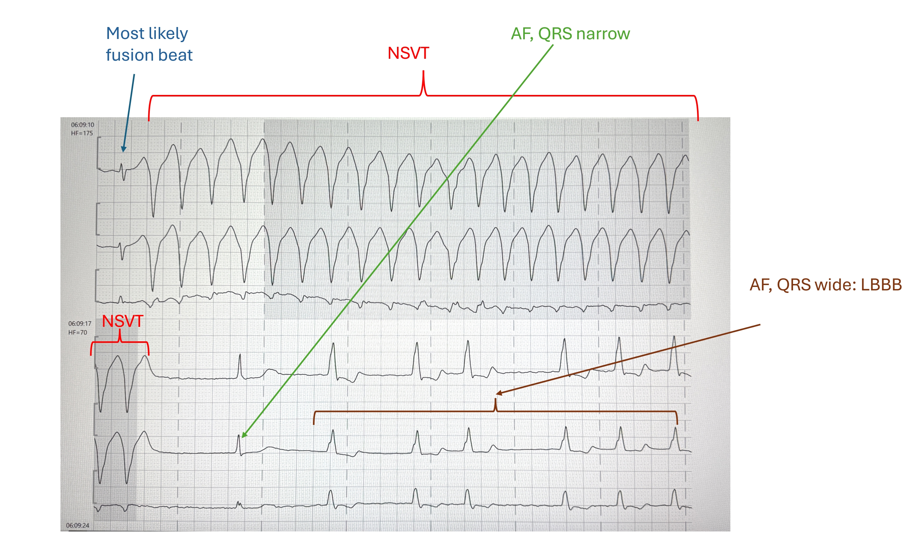
Many people are considerably unsettled by ECG recordings from smart watches. However, smartwatch ECGs can be helpful in the diagnosis of paroxysmal atrial fibrillation. Here. you can see an example. It is a 1-channel ECG that corresponds to lead I. Initially there is an irregular fast pulse without P waves, which corresponds to a tachyarrhythmia in atrial fibrillation. At the end of the first line, after a very short pause, there is sinus rhythm. In the third line you see a short SVT over 3 beats, then SR again. The QRS complex is widened to over 120 ms.

Dr A Röschl's picture

Atypical Atrial Flutter (From the Left Atrium)

Why is this atypical atrial flutter from the left atrium?

Dr A Röschl's picture

Ventricular Tachycardia

There are several differential diagnoses in the presence of broad-complex tachycardia. The most common cause of wide-complex tachycardia is ventricular tachycardia. In 2nd place is sinus tachycardia/supraventricular tachycardia with aberrant conduction or preexisting bundle branch block.

Dr A Röschl's picture

Right Bundle Branch Block

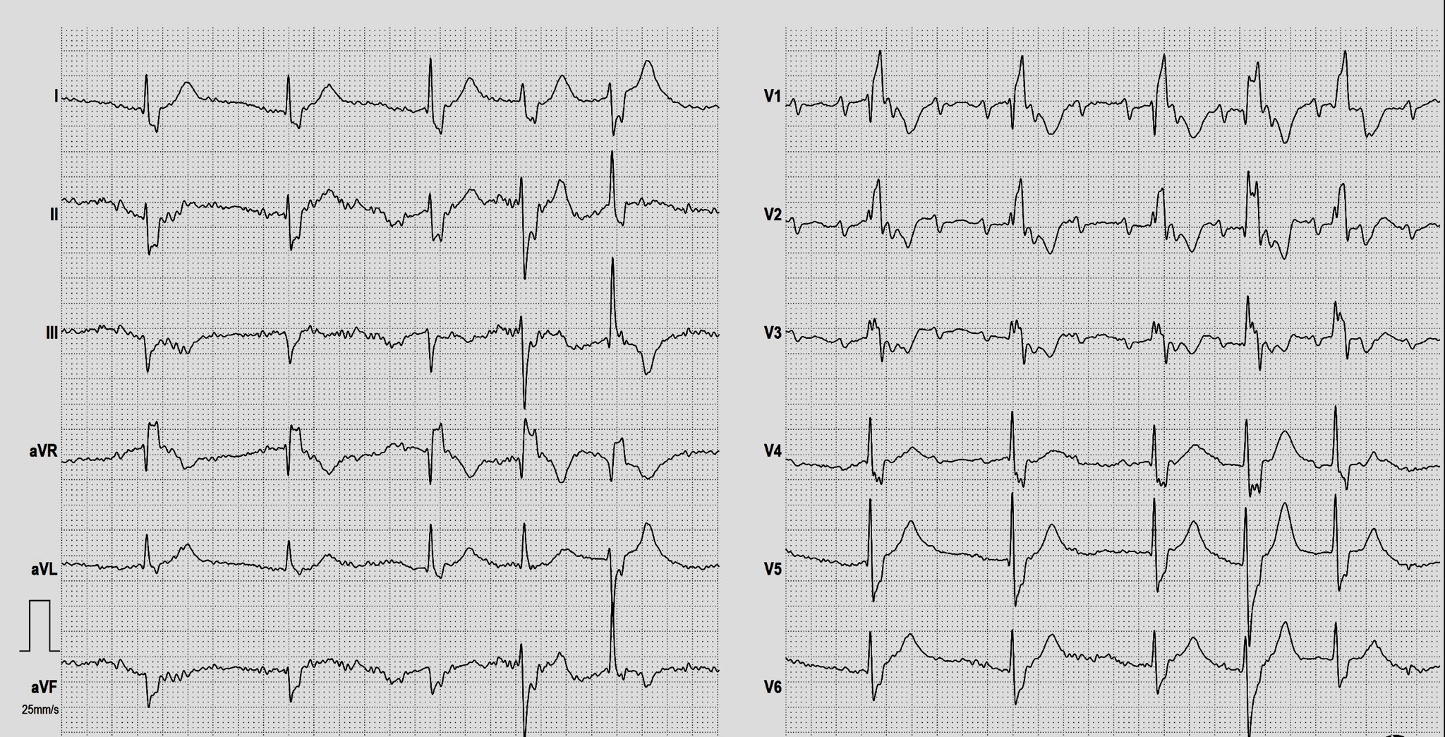
Why is this a right bundle branch block? We see a SR with broad QRS complexes (more than 120 ms). These are positive in the rightward leads III and especially in V1 and V2. In the more leftward leads I, aVL, V4-V6 there are clear S waves. This is a typical RBBB pattern.

Dr A Röschl's picture

Left Bundle Branch Block

Why is this a left bundle branch block? We see a SR with broad QRS complexes (more than 120 ms). These are positive in the leftward leads I and aVL and in V5 and V6. In the more rightward leads III, aVR, and V1-V3, the QRS complexes are predominantly negative with deep S waves. This is a typical LBBB pattern.

Dr A Röschl's picture

AVNRT TYPICAL FORM

18-year-old male; palpitations lasting hours, beginning at the age of 10. What arrhythmia is present? Let's first consider the heart rate: with a heart rate of 194 beats/min, the heart rate is too low for atrial flutter (1:1) (except in patients who have been pre-treated with medication), and the rate would be unusually high for atrial flutter with 2:1 conduction. Due to the regularity of the heart rhythm, atrial fibrillation can also be ruled out. This leaves atrial tachycardia, junctional tachycardia, AVNRT, and AVRT as possibilities.

Dr A Röschl's picture

Nonconducted PACs

A common cause of pauses and bradycardia are non-conducted PACs, which generally do not require treatment. Therefore, it is important to differentiate between pauses or bradycardia that require treatment.

Dr A Röschl's picture

Polymorphic ventricular tachycardia during a stress EKG. What is the most likely cause?

This EKG shows a sinus rhythm with ventricular bigeminy and retrograde conduction leading to retrograde depolarization of the sinus node, resulting in a longer pause (sinus node reset).
Then, a polymorphic ventricular tachycardia occurs over 7 beats. The QT interval of the sinus beats does not appear prolonged, thus ruling out Torsades de Pointes tachycardia. The most likely cause of this type of polymorphic ventricular tachycardia during a stress EKG is cardiac ischemia/coronary artery disease.

Dr A Röschl's picture

SICK SINUS SYNDROME

This EKG shows the classic features of sick sinus syndrome. Initially, there is an accelerated atrial rhythm/atrial tachycardia. After a pre-automatic pause of 2609 ms, a ventricular premature beat occurs, followed by a junctional escape rhythm. Pre-automatic pause is a pause after a tachycardia and before an automatic rhythm, like sinus rhythm or, in this case, junctional escape rhythm. Therefore, there is an alternation between tachycardic phases and very bradycardic rhythms, the classic bradycardia/tachycardia syndrome. Paper speed is 12.5 mm/sec.

Dr A Röschl's picture

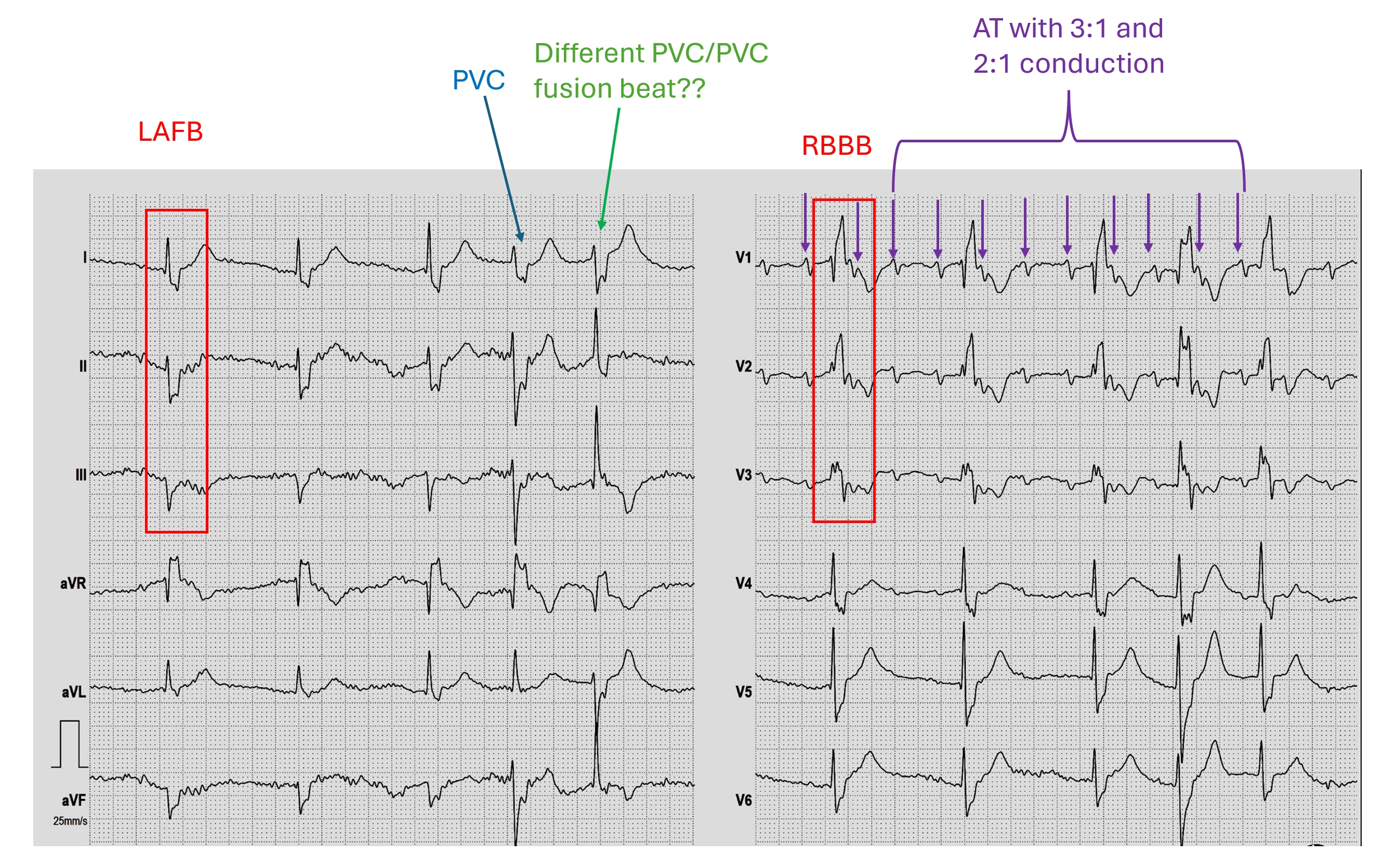
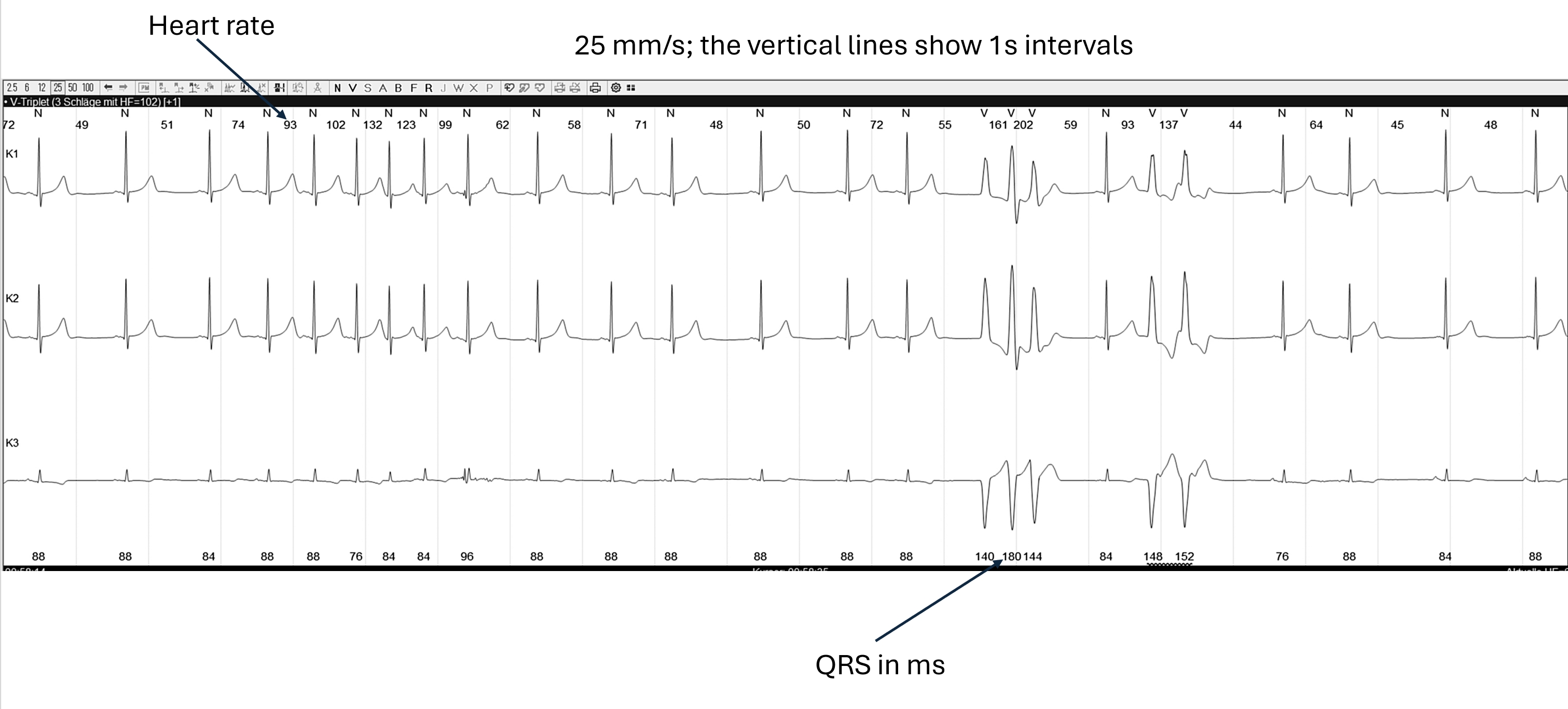
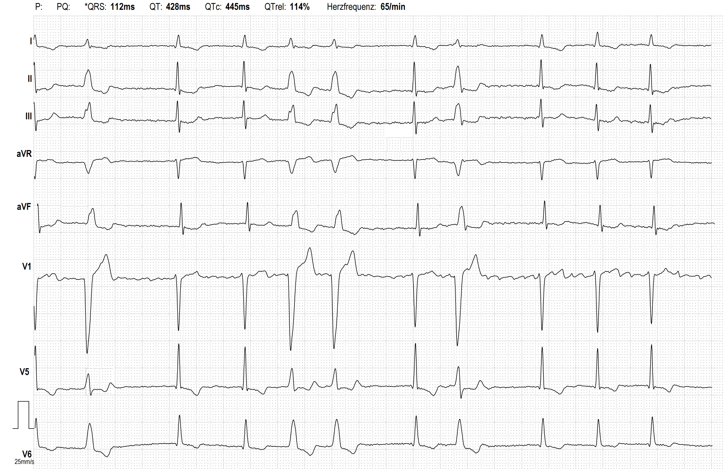
Impending Trifascicular AV Block

Here we see the EKG of a 63-year-old man with CAD without relevant coronary stenosis. He complains of slightly reduced performance, but no other symptoms. The ECG shows the following changes:

Pages

All our content is FREE & COPYRIGHT FREE for non-commercial use

Please be courteous and leave any watermark or author attribution on content you reproduce.